 Explore
 Validate
 Learn
Learn Western blot
Antibody data
- Antibody Data
- Antigen structure
- References [0]
- Comments [0]
- Validations
- Western blot [3]
- Immunohistochemistry [1]
- Chromatin Immunoprecipitation [1]
Submit
Validation data
Reference
Comment
Report error
- Product number
- PA1-38675 - Provider product page

- Provider
- Invitrogen Antibodies
- Product name
- STAT6 Polyclonal Antibody
- Antibody type
- Polyclonal
- Antigen
- Synthetic peptide
- Description
- This antibody is predicted to react with canine, mouse, porcine, rabbit based on sequence homology. Heat-mediated antigen retrieval is recommended prior to staining, using a 10mM citrate buffer, pH 6.0, for 10 minutes followed by cooling at room temperature for 20 min. Following antigen retrieval, incubate samples with primary antibody for 10 min at room temperature. A suggested positive control is placenta or tonsil tissue.
- Reactivity
- Human
- Host
- Rabbit
- Isotype
- IgG
- Vial size
- 1 mL
- Concentration
- 0.28 mg/mL
- Storage
- -20° C, Avoid Freeze/Thaw Cycles
No comments: Submit comment
Supportive validation
- Submitted by
- Invitrogen Antibodies (provider)
- Main image

- Experimental details
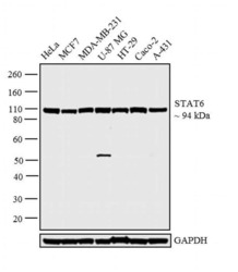
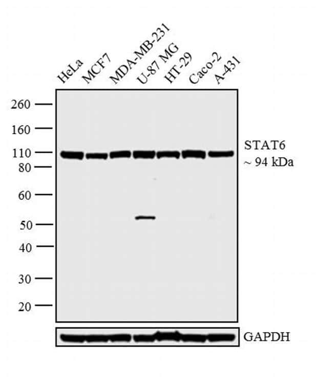
- Western blot analysis was performed on whole cell extracts (30 µg lysate) of HeLa (Lane 1), MCF7 (Lane 2), MDA-MB-231 (Lane 3), U-87 MG (Lane 4), HT-29 (Lane 5), Caco-2 (Lane 6) and A-431 (Lane 7). The blot was probed with Anti-STAT6 Polyclonal Antibody (Product # PA5-32603, 1:500 dilution) and detected by chemiluminescence using Goat anti-Rabbit IgG (H+L) Superclonal™ Secondary Antibody, HRP conjugate (Product # A27036, 0.25 µg/mL, 1:4000 dilution). A 94 kDa band corresponding to STAT6 was observed across the cell lines tested.

- Submitted by
- Invitrogen Antibodies (provider)
- Main image

- Experimental details
- Western blot analysis was performed on whole cell extracts (30 µg lysate) of HeLa (Lane 1), MCF7 (Lane 2), MDA-MB-231 (Lane 3), U-87 MG (Lane 4), HT-29 (Lane 5), Caco-2 (Lane 6) and A-431 (Lane 7). The blot was probed with Anti-STAT6 Polyclonal Antibody (Product # PA5-32603, 1:500 dilution) and detected by chemiluminescence using Goat anti-Rabbit IgG (H+L) Superclonal™ Secondary Antibody, HRP conjugate (Product # A27036, 0.25 µg/mL, 1:4000 dilution). A 94 kDa band corresponding to STAT6 was observed across the cell lines tested.

- Submitted by
- Invitrogen Antibodies (provider)
- Main image
- Experimental details
- Western blot analysis was performed on whole cell extracts (30 µg lysate) of HeLa (Lane 1), MCF7 (Lane 2), MDA-MB-231 (Lane 3), U-87 MG (Lane 4), HT-29 (Lane 5), Caco-2 (Lane 6) and A-431 (Lane 7). The blot was probed with Anti-STAT6 Polyclonal Antibody (Product # PA5-32603, 1:500 dilution) and detected by chemiluminescence using Goat anti-Rabbit IgG (H+L) Superclonal™ Secondary Antibody, HRP conjugate (Product # A27036, 0.25 µg/mL, 1:4000 dilution). A 94 kDa band corresponding to STAT6 was observed across the cell lines tested.
Supportive validation
- Submitted by
- Invitrogen Antibodies (provider)
- Main image

- Experimental details
- Immunohistochemical analysis of STAT6 using anti-STAT6 Polyclonal Antibody (Product # PA5-32603) in Placenta Tissue. The recommened dilution for this antibody in immunohistochemistry applications is 1:150.

Supportive validation
- Submitted by
- Invitrogen Antibodies (provider)
- Main image

- Experimental details
- Enrichment of endogenous STAT6 protein at specific gene loci using Anti-STAT6 Antibody: Chromatin Immunoprecipitation (ChIP) was performed using Anti-STAT6 Rabbit Polyclonal Antibody (Product # PA5-32603, 12 µl) on sheared chromatin from 2 million HeLa cells treated with IL4 (overnight serum-starvation followed by 100 ng/mL IL4 for 30 min) using the MAGnify ChIP system kit (Product # 49-2024). Normal Rabbit IgG was used as a negative IP control. The purified DNA was analyzed by qPCR with PCR primer pairs over MS4A1 and DMD promoters, DMD intron 2 (active) and SAT2 satellite repeats (inactive). Data is presented as fold enrichment of the antibody signal versus the negative control IgG using the comparative CT method.
