Explore
 Validate
 Learn
LearnPA5-17564
antibody from Invitrogen Antibodies
Targeting: ACACA
ACAC, ACC, ACC1
 Western blot Immunocytochemistry
 Immunoprecipitation Immunohistochemistry Flow cytometry Other assay
Antibody data
- Antibody Data
- Antigen structure
- References [4]
- Comments [0]
- Validations
- Western blot [2]
- Immunocytochemistry [1]
- Immunohistochemistry [1]
- Other assay [2]
Submit
Validation data
Reference
Comment
Report error
- Product number
- PA5-17564 - Provider product page

- Provider
- Invitrogen Antibodies
- Product name
- Acetyl-CoA Carboxylase Polyclonal Antibody
- Antibody type
- Polyclonal
- Antigen
- Synthetic peptide
- Description
- It is not recommended to aliquot this antibody.
- Reactivity
- Human, Mouse, Rat, Bovine
- Host
- Rabbit
- Isotype
- IgG
- Vial size
- 100 µL
- Concentration
- 17 µg/mL
- Storage
- -20°C
Submitted references LncEDCH1 improves mitochondrial function to reduce muscle atrophy by interacting with SERCA2.
Berberine (BBR) Attenuated Palmitic Acid (PA)-Induced Lipotoxicity in Human HK-2 Cells by Promoting Peroxisome Proliferator-Activated Receptor α (PPAR-α).
Acetyl-CoA carboxylase 1-dependent lipogenesis promotes autophagy downstream of AMPK.
Green tea polyphenols alter lipid metabolism in the livers of broiler chickens through increased phosphorylation of AMP-activated protein kinase.
Cai B, Ma M, Zhang J, Wang Z, Kong S, Zhou Z, Lian L, Zhang J, Li J, Wang Y, Li H, Zhang X, Nie Q
Molecular therapy. Nucleic acids 2022 Mar 8;27:319-334
Molecular therapy. Nucleic acids 2022 Mar 8;27:319-334
Berberine (BBR) Attenuated Palmitic Acid (PA)-Induced Lipotoxicity in Human HK-2 Cells by Promoting Peroxisome Proliferator-Activated Receptor α (PPAR-α).
Wu Y, Chen F, Huang X, Zhang R, Yu Z, Chen Z, Liu J
Medical science monitor : international medical journal of experimental and clinical research 2019 Oct 14;25:7702-7708
Medical science monitor : international medical journal of experimental and clinical research 2019 Oct 14;25:7702-7708
Acetyl-CoA carboxylase 1-dependent lipogenesis promotes autophagy downstream of AMPK.
Gross AS, Zimmermann A, Pendl T, Schroeder S, Schoenlechner H, Knittelfelder O, Lamplmayr L, Santiso A, Aufschnaiter A, Waltenstorfer D, Ortonobes Lara S, Stryeck S, Kast C, Ruckenstuhl C, Hofer SJ, Michelitsch B, Woelflingseder M, Müller R, Carmona-Gutierrez D, Madl T, Büttner S, Fröhlich KU, Shevchenko A, Eisenberg T
The Journal of biological chemistry 2019 Aug 9;294(32):12020-12039
The Journal of biological chemistry 2019 Aug 9;294(32):12020-12039
Green tea polyphenols alter lipid metabolism in the livers of broiler chickens through increased phosphorylation of AMP-activated protein kinase.
Huang J, Zhou Y, Wan B, Wang Q, Wan X
PloS one 2017;12(10):e0187061
PloS one 2017;12(10):e0187061
No comments: Submit comment
Supportive validation
- Submitted by
- Invitrogen Antibodies (provider)
- Main image

- Experimental details
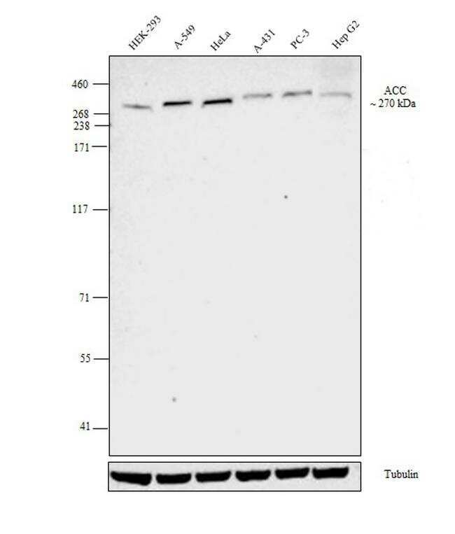
- Western blot analysis was performed on membrane enriched extracts (30 µg lysate) of HEK-293 (Lane 1), A549 (Lane 2), HeLa (Lane 3), A-431 (Lane 4), PC-3 (Lane 5), Hep G2(Lane 6). The blot was probed with Acetyl-CoA Carboxylase Polyclonal Antibody(Product # PA5-17564, 1:1000) and detected by chemiluminescence using Goat anti-Rabbit IgG (H+L) Superclonal™ Secondary Antibody, HRP conjugate (Product # A27036, 0.25 µg/ml, 1:4000 dilution). A band at ~270 kDa corresponding to Acetyl-CoA was observed across all the cell lines tested.
- Submitted by
- Invitrogen Antibodies (provider)
- Main image
- Experimental details
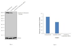
- Knockdown of AcetylCoA Carboxylase was achieved by transfecting HeLa cells with AcetylCoA Carboxylase specific siRNAs (Silencer® select Product # s882, s883). Western blot analysis (Fig. a) was performed using whole cell extracts from the AcetylCoA Carboxylase knockdown cells (lane 3), non-specific scrambled siRNA transfected cells (lane 2) and untransfected cells (lane 1). The blots were probed with AcetylCoA Carboxylase Polyclonal Antibody (Product # MA5-17564, 1:1000 dilution) and Goat anti-Rabbit IgG (H+L) Superclonal™ Secondary Antibody, HRP conjugate (Product # A27036, 0.25 µg/ml, 1:4000 dilution). Densitometric analysis of this western blot is shown in histogram (Fig. b). Decrease in signal upon siRNA mediated knock down confirms that antibody is specific to AcetylCoA Carboxylase.

Supportive validation
- Submitted by
- Invitrogen Antibodies (provider)
- Main image

- Experimental details
- Immunofluorescent analysis of Acetyl-CoA Carboxylase in A431 cells using an Acetyl-CoA Carboxylase polyclonal antibody (Product # PA5-17564) (A) compared to an isotype control (B) showing cytoplasmic staining.

Supportive validation
- Submitted by
- Invitrogen Antibodies (provider)
- Main image

- Experimental details
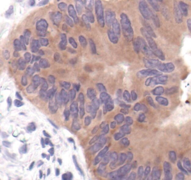
- Immunohistochemical analysis of Acetyl-CoA Carboxylase in paraffin-embedded human colon carcinoma using an Acetyl-CoA Carboxylase polyclonal antibody (Product # PA5-17564) in the presence of control peptide (left) or Acetyl-CoA Carboxylase blocking peptide (right).
Supportive validation
- Submitted by
- Invitrogen Antibodies (provider)
- Main image

- Experimental details
- Figure 1. Acc1 activity correlates with autophagy levels. WT and acc1-S1157A mutant ( acc1 S/A ) yeast cells were aged until the indicated time points on 2% glucose minimal medium. SorA or the solvent DMSO was applied 6 h after inoculation where indicated. A , schematic overview of the Acc1-regulated metabolic pathway. Acc1 activity can be modulated by SorA treatment (inhibition, red color ) or the S1157A point mutation (activation, blue color ). B , flow cytometric quantification of neutral lipids after BODIPY staining in a time course experiment. Relative fluorescence units were normalized to the WT at 24 h ( n = 4). C and D , representative immunoblots ( C ) and quantification of free GFP/GAPDH ( D ), indicating autophagic flux ( GFP liberation assay ) of GFP-Atg8-expressing cells after 2 days of aging. Blots were probed with Acc1-, GFP-, or GAPDH-specific antibodies, and immunoblot signals were normalized to the WT. ( n = 4). E and F , flow cytometric quantification of neutral lipids after BODIPY staining in a time course experiment ( E ) or after 24 h of incubation (day 1 of aging) ( F ). The arrow in E indicates the time of SorA application. Relative fluorescence units were normalized to the WT control at 24 h ( n = 4 in E ; n = 7 in F ). G and H , representative immunoblots ( G ) and densitometric quantification of free GFP/GAPDH ( H , left ) or full-length GFP-Atg8/GAPDH levels ( H , right ) of GFP-Atg8-expressing cells at the indicated age ( G ) or after 2 days of ag

- Submitted by
- Invitrogen Antibodies (provider)
- Main image
- Experimental details
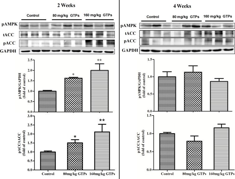
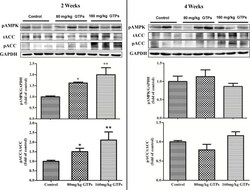
- Fig 4 Phosphorylation of hepatic AMPKalpha and ACACA in broilers treated with green tea polyphenols. Broilers were treated with vehicle (distilled water), 80 mg/kg GTPs, or 160 mg/kg GTPs for 2 or 4 weeks. Western blots were probed with antibodies targeting phosphorylated version of ACACA, acetyl-CoA carboxylase and AMPK, AMP-activated protein kinase or unphosphorylated version of ACACA and GAPDH, glyceraldehyde-3-phosphate dehydrogenase. Values are represented as the means +- SEM (n = 6); statistical significance was determined by ANOVA with Duncan's multiple-range test, with * p < 0.05 and ** p < 0.01 compared with control.