 Explore
 Validate
 Learn
Learn Immunocytochemistry
 Immunoprecipitation
Antibody data
- Antibody Data
- Antigen structure
- References [16]
- Comments [0]
- Validations
- Immunocytochemistry [2]
- Immunohistochemistry [1]
- Other assay [1]
Submit
Validation data
Reference
Comment
Report error
- Product number
- MA5-12998 - Provider product page

- Provider
- Invitrogen Antibodies
- Product name
- ErbB2 (HER-2) Monoclonal Antibody (N12)
- Antibody type
- Monoclonal
- Antigen
- Other
- Description
- MA5-12998 targets HER-2 in ICC/IF, IHC (F), IHC (P) and IM applications and shows reactivity with Human samples. The MA5-12998 immunogen is intact SKBR-3 breast cancer cells.
- Reactivity
- Human
- Host
- Mouse
- Isotype
- IgG
- Antibody clone number
- N12
- Vial size
- 500 µL
- Concentration
- 0.2 mg/mL
- Storage
- 4° C
Submitted references Anks1a regulates COPII-mediated anterograde transport of receptor tyrosine kinases critical for tumorigenesis.
Truncated ErbB2 expressed in tumor cell nuclei contributes to acquired therapeutic resistance to ErbB2 kinase inhibitors.
Quantitative assays for the measurement of HER1-HER2 heterodimerization and phosphorylation in cell lines and breast tumors: applications for diagnostics and targeted drug mechanism of action.
Analytical Validation of a Highly Quantitative, Sensitive, Accurate, and Reproducible Assay (HERmark) for the Measurement of HER2 Total Protein and HER2 Homodimers in FFPE Breast Cancer Tumor Specimens.
Silica-gold nanoshells as potential intraoperative molecular probes for HER2-overexpression in ex vivo breast tissue using near-infrared reflectance confocal microscopy.
Impact of common epidermal growth factor receptor and HER2 variants on receptor activity and inhibition by lapatinib.
Enhanced multi-spectral imaging of live breast cancer cells using immunotargeted gold nanoshells and two-photon excitation microscopy.
Delineation of molecular mechanisms of sensitivity to lapatinib in breast cancer cell lines using global gene expression profiles.
Immunonanoshells for targeted photothermal ablation of tumor cells.
Immunonanoshells for targeted photothermal ablation of tumor cells.
Tumor cell expression of HLA-DM associates with a Th1 profile and predicts improved survival in breast carcinoma patients.
Isolation of scFvs to in vitro produced extracellular domains of EGFR family members.
Binding at and transactivation of the COX-2 promoter by nuclear tyrosine kinase receptor ErbB-2.
Truncated ErbB2 receptor (p95ErbB2) is regulated by heregulin through heterodimer formation with ErbB3 yet remains sensitive to the dual EGFR/ErbB2 kinase inhibitor GW572016.
C-erbB2 oncoprotein and its soluble ectodomain: a new potential tumor marker for prognosis early detection and monitoring patients undergoing Herceptin treatment.
C-erbB2 oncoprotein and its soluble ectodomain: a new potential tumor marker for prognosis early detection and monitoring patients undergoing Herceptin treatment.
Lee H, Noh H, Mun J, Gu C, Sever S, Park S
Nature communications 2016 Sep 13;7:12799
Nature communications 2016 Sep 13;7:12799
Truncated ErbB2 expressed in tumor cell nuclei contributes to acquired therapeutic resistance to ErbB2 kinase inhibitors.
Xia W, Liu Z, Zong R, Liu L, Zhao S, Bacus SS, Mao Y, He J, Wulfkuhle JD, Petricoin EF 3rd, Osada T, Yang XY, Hartman ZC, Clay TM, Blackwell KL, Lyerly HK, Spector NL
Molecular cancer therapeutics 2011 Aug;10(8):1367-74
Molecular cancer therapeutics 2011 Aug;10(8):1367-74
Quantitative assays for the measurement of HER1-HER2 heterodimerization and phosphorylation in cell lines and breast tumors: applications for diagnostics and targeted drug mechanism of action.
DeFazio-Eli L, Strommen K, Dao-Pick T, Parry G, Goodman L, Winslow J
Breast cancer research : BCR 2011 Apr 15;13(2):R44
Breast cancer research : BCR 2011 Apr 15;13(2):R44
Analytical Validation of a Highly Quantitative, Sensitive, Accurate, and Reproducible Assay (HERmark) for the Measurement of HER2 Total Protein and HER2 Homodimers in FFPE Breast Cancer Tumor Specimens.
Larson JS, Goodman LJ, Tan Y, Defazio-Eli L, Paquet AC, Cook JW, Rivera A, Frankson K, Bose J, Chen L, Cheung J, Shi Y, Irwin S, Kiss LD, Huang W, Utter S, Sherwood T, Bates M, Weidler J, Parry G, Winslow J, Petropoulos CJ, Whitcomb JM
Pathology research international 2010 Jun 28;2010:814176
Pathology research international 2010 Jun 28;2010:814176
Silica-gold nanoshells as potential intraoperative molecular probes for HER2-overexpression in ex vivo breast tissue using near-infrared reflectance confocal microscopy.
Bickford LR, Agollah G, Drezek R, Yu TK
Breast cancer research and treatment 2010 Apr;120(3):547-55
Breast cancer research and treatment 2010 Apr;120(3):547-55
Impact of common epidermal growth factor receptor and HER2 variants on receptor activity and inhibition by lapatinib.
Gilmer TM, Cable L, Alligood K, Rusnak D, Spehar G, Gallagher KT, Woldu E, Carter HL, Truesdale AT, Shewchuk L, Wood ER
Cancer research 2008 Jan 15;68(2):571-9
Cancer research 2008 Jan 15;68(2):571-9
Enhanced multi-spectral imaging of live breast cancer cells using immunotargeted gold nanoshells and two-photon excitation microscopy.
Bickford L, Sun J, Fu K, Lewinski N, Nammalvar V, Chang J, Drezek R
Nanotechnology 2008 Aug 6;19(31):315102
Nanotechnology 2008 Aug 6;19(31):315102
Delineation of molecular mechanisms of sensitivity to lapatinib in breast cancer cell lines using global gene expression profiles.
Hegde PS, Rusnak D, Bertiaux M, Alligood K, Strum J, Gagnon R, Gilmer TM
Molecular cancer therapeutics 2007 May;6(5):1629-40
Molecular cancer therapeutics 2007 May;6(5):1629-40
Immunonanoshells for targeted photothermal ablation of tumor cells.
Lowery AR, Gobin AM, Day ES, Halas NJ, West JL
International journal of nanomedicine 2006;1(2):149-54
International journal of nanomedicine 2006;1(2):149-54
Immunonanoshells for targeted photothermal ablation of tumor cells.
Lowery AR, Gobin AM, Day ES, Halas NJ, West JL
International journal of nanomedicine 2006;1(2):149-54
International journal of nanomedicine 2006;1(2):149-54
Tumor cell expression of HLA-DM associates with a Th1 profile and predicts improved survival in breast carcinoma patients.
Oldford SA, Robb JD, Codner D, Gadag V, Watson PH, Drover S
International immunology 2006 Nov;18(11):1591-602
International immunology 2006 Nov;18(11):1591-602
Isolation of scFvs to in vitro produced extracellular domains of EGFR family members.
Horak E, Heitner T, Robinson MK, Simmons HH, Garrison J, Russeva M, Furmanova P, Lou J, Zhou Y, Yuan QA, Weiner LM, Adams GP, Marks JD
Cancer biotherapy & radiopharmaceuticals 2005 Dec;20(6):603-13
Cancer biotherapy & radiopharmaceuticals 2005 Dec;20(6):603-13
Binding at and transactivation of the COX-2 promoter by nuclear tyrosine kinase receptor ErbB-2.
Wang SC, Lien HC, Xia W, Chen IF, Lo HW, Wang Z, Ali-Seyed M, Lee DF, Bartholomeusz G, Ou-Yang F, Giri DK, Hung MC
Cancer cell 2004 Sep;6(3):251-61
Cancer cell 2004 Sep;6(3):251-61
Truncated ErbB2 receptor (p95ErbB2) is regulated by heregulin through heterodimer formation with ErbB3 yet remains sensitive to the dual EGFR/ErbB2 kinase inhibitor GW572016.
Xia W, Liu LH, Ho P, Spector NL
Oncogene 2004 Jan 22;23(3):646-53
Oncogene 2004 Jan 22;23(3):646-53
C-erbB2 oncoprotein and its soluble ectodomain: a new potential tumor marker for prognosis early detection and monitoring patients undergoing Herceptin treatment.
Wu JT
Clinica chimica acta; international journal of clinical chemistry 2002 Aug;322(1-2):11-9
Clinica chimica acta; international journal of clinical chemistry 2002 Aug;322(1-2):11-9
No comments: Submit comment
Supportive validation
- Submitted by
- Invitrogen Antibodies (provider)
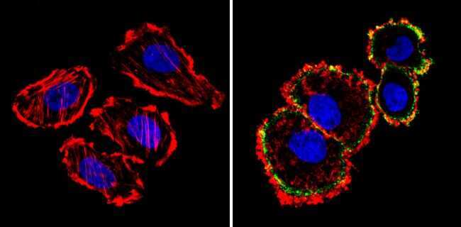
- Main image

- Experimental details
- Immunofluorescent analysis of HER-2 (green) showing staining in the membrane of SK-BR-3 cells (right) compared to a negative control without primary antibody (left). Formalin-fixed cells were permeabilized with 0.1% Triton X-100 in TBS for 5-10 minutes and blocked with 3% BSA-PBS for 30 minutes at room temperature. Cells were probed with a HER-2 monoclonal antibody (Product # MA5-12998) in 3% BSA-PBS at a dilution of 1:100 and incubated overnight at 4 ºC in a humidified chamber. Cells were washed with PBST and incubated with a DyLight-conjugated secondary antibody in PBS at room temperature in the dark. F-actin (red) was stained with a fluorescent red phalloidin and nuclei (blue) were stained with Hoechst or DAPI. Images were taken at a magnification of 60x.
- Submitted by
- Invitrogen Antibodies (provider)
- Main image
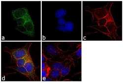
- Experimental details
- Immunofluorescence analysis of ErbB2 was performed using 90% confluent log phase MCF7 cells. The cells were fixed with 4% paraformaldehyde for 10 minutes, permeabilized with 0.1% Triton™ X-100 for 10 minutes, and blocked with 1% BSA for 1 hour at room temperature. The cells were labeled with ErbB2 (N12) Mouse Monoclonal Antibody (Product # MA5-12998) at 1:250 dilution in 0.1% BSA and incubated for 3 hours at room temperature and then labeled with Goat anti-Mouse IgG (H+L) Superclonal™ Secondary Antibody, Alexa Fluor® 488 conjugate (Product # A28175) at a dilution of 1:2000 for 45 minutes at room temperature (Panel a: green). Nuclei (Panel b: blue) were stained with SlowFade® Gold Antifade Mountant with DAPI (Product # S36938). F-actin (Panel c: red) was stained with Rhodamine Phalloidin (Product # R415, 1:300). Panel d represents the merged image showing membranous and cytoplasmic localization. Panel e shows the no primary antibody control. The images were captured at 60X magnification.

Supportive validation
- Submitted by
- Invitrogen Antibodies (provider)
- Main image

- Experimental details
- Immunohistochemistry analysis of HER-2 showing positive staining in the cytoplasm, nucleus and membrane of paraffin-treated Human breast carcinoma (right) compared with a negative control in the absence of primary antibody (left). To expose target proteins, antigen retrieval method was performed using 10mM sodium citrate (pH 6.0) microwaved for 8-15 min. Following antigen retrieval, tissues were blocked in 3% H2O2-methanol for 15 min at room temperature, washed with ddH2O and PBS, and then probed with a HER-2 monoclonal antibody (Product # MA5-12998) diluted by 3% BSA-PBS at a dilution of 1:100 overnight at 4°C in a humidified chamber. Tissues were washed extensively PBST and detection was performed using an HRP-conjugated secondary antibody followed by colorimetric detection using a DAB kit. Tissues were counterstained with hematoxylin and dehydrated with ethanol and xylene to prep for mounting.

Supportive validation
- Submitted by
- Invitrogen Antibodies (provider)
- Main image

- Experimental details
- Figure 5 Anks1a influences breast tumorigenesis by regulating the ER export of EphA2-ErbB2 complexes. ( a ) Anks1a +/- female mice were crossed with Anks1a +/- ; MMTV-Neu male mice to obtain female mice with the indicated genotypes. All experiments monitoring breast tumour formation were carried out in mice of the FVB genetic background ( n =16, 29, 14 per genotype). ** P
