Explore
 Validate
 Learn
Learn Immunocytochemistry
 Immunoprecipitation
Antibody data
- Antibody Data
- Antigen structure
- References [7]
- Comments [0]
- Validations
- Immunocytochemistry [1]
Submit
Validation data
Reference
Comment
Report error
- Product number
- MA5-13679 - Provider product page

- Provider
- Invitrogen Antibodies
- Product name
- ErbB2 (HER-2) Monoclonal Antibody (L26)
- Antibody type
- Monoclonal
- Antigen
- Recombinant full-length protein
- Description
- MA5-13679 targets HER-2 in immunoprecipitation applications and shows reactivity with Human samples. The MA5-13679 immunogen is extracellular domain of recombinant human c-erbB-2/HER-2 oncoprotein.
- Reactivity
- Human
- Host
- Mouse
- Isotype
- IgG
- Antibody clone number
- L26
- Vial size
- 500 µL
- Concentration
- 0.2 mg/mL
- Storage
- -20° C, Avoid Freeze/Thaw Cycles
Submitted references Association between growth factor heregulin1α and receptors in growth of ovarian cancer cell line with high potentiality of peritoneal dissemination.
BCL6 positively regulates AID and germinal center gene expression via repression of miR-155.
Lymphoepithelioma-like carcinoma of the ovary.
Identification of a morphometrical parameter that predicts the response to splenectomy in patients with idiopathic thrombocytopenic purpura.
A chimeric multi-human epidermal growth factor receptor-2 B cell epitope peptide vaccine mediates superior antitumor responses.
A chimeric multi-human epidermal growth factor receptor-2 B cell epitope peptide vaccine mediates superior antitumor responses.
Identification of epitope regions recognized by tumor inhibitory and stimulatory anti-ErbB-2 monoclonal antibodies: implications for vaccine design.
Nishiyama H, Soeda S, Watanabe T, Fujimori K
Fukushima journal of medical science 2012;58(1):22-32
Fukushima journal of medical science 2012;58(1):22-32
BCL6 positively regulates AID and germinal center gene expression via repression of miR-155.
Basso K, Schneider C, Shen Q, Holmes AB, Setty M, Leslie C, Dalla-Favera R
The Journal of experimental medicine 2012 Dec 17;209(13):2455-65
The Journal of experimental medicine 2012 Dec 17;209(13):2455-65
Lymphoepithelioma-like carcinoma of the ovary.
Ambrosio MR, Rocca BJ, Onorati M, Mourmouras V, Mastrogiulio MG, Crispino S, Liberatore C, Santopietro R
International journal of surgical pathology 2011 Aug;19(4):514-7
International journal of surgical pathology 2011 Aug;19(4):514-7
Identification of a morphometrical parameter that predicts the response to splenectomy in patients with idiopathic thrombocytopenic purpura.
Bakkaloglu H, Dinccag A, Yanar H, Tunca F, Dogan O, Cermik H, Kucukkaya R
The Tohoku journal of experimental medicine 2006 Sep;210(1):49-55
The Tohoku journal of experimental medicine 2006 Sep;210(1):49-55
A chimeric multi-human epidermal growth factor receptor-2 B cell epitope peptide vaccine mediates superior antitumor responses.
Dakappagari NK, Pyles J, Parihar R, Carson WE, Young DC, Kaumaya PT
Journal of immunology (Baltimore, Md. : 1950) 2003 Apr 15;170(8):4242-53
Journal of immunology (Baltimore, Md. : 1950) 2003 Apr 15;170(8):4242-53
Identification of epitope regions recognized by tumor inhibitory and stimulatory anti-ErbB-2 monoclonal antibodies: implications for vaccine design.
Yip YL, Smith G, Koch J, Dübel S, Ward RL
Journal of immunology (Baltimore, Md. : 1950) 2001 Apr 15;166(8):5271-8
Journal of immunology (Baltimore, Md. : 1950) 2001 Apr 15;166(8):5271-8
No comments: Submit comment
Supportive validation
- Submitted by
- Invitrogen Antibodies (provider)
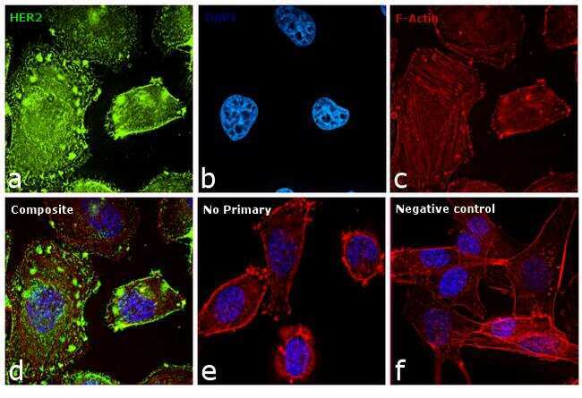
- Main image
- Experimental details
- Immunofluorescence analysis of Her2 was performed using 70% confluent log phase SK-BR-3 cells. The cells were fixed with 4% paraformaldehyde for 10 minutes, permeabilized with 0.1% Triton™ X-100 for 10 minutes, and blocked with 1% BSA for 1 hour at room temperature. The cells were labeled with Her2 Mouse monoclonal Antibody (Product # MA5-13679) at 5 µg/mL in 0.1% BSA and incubated overnight at 4 degree Celsius and then labeled with Goat anti-Mouse IgG (H+L) Superclonal™ Secondary Antibody, Alexa Fluor® 488 conjugate (Product # A28175) at a dilution of 1:2000 for 45 minutes at room temperature (Panel a: green). Nuclei (Panel b: blue) were stained with SlowFade® Gold Antifade Mountant with DAPI (Product # S36938). F-actin (Panel c: red) was stained with Rhodamine Phalloidin (Product # R415, 1:300). Panel d represents the merged image showing membranous and cytoplasmic localization. Panel f represents MDAMB-231 cells as negative controls, showing no Her2 staining. Panel e represents control cells with no primary antibody to assess background. The images were captured at 60X magnification.