Explore
 Validate
 Learn
Learn Western blot
Antibody data
- Antibody Data
- Antigen structure
- References [0]
- Comments [0]
- Validations
- Western blot [1]
- Immunocytochemistry [2]
Submit
Validation data
Reference
Comment
Report error
- Product number
- PA5-37565 - Provider product page

- Provider
- Invitrogen Antibodies
- Product name
- Phospho-ErbB2 (HER-2) (Tyr1221, Tyr1222) Polyclonal Antibody
- Antibody type
- Polyclonal
- Antigen
- Synthetic peptide
- Description
- A suggested positive control for Western blot is MDA cells; suggested positive control for ICC/IF is MCF-7 cells.
- Reactivity
- Human
- Host
- Rabbit
- Isotype
- IgG
- Vial size
- 100 µL
- Concentration
- 1 mg/mL
- Storage
- -20°C
No comments: Submit comment
Supportive validation
- Submitted by
- Invitrogen Antibodies (provider)
- Main image
- Experimental details
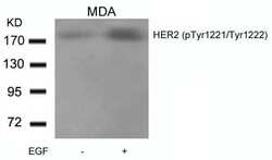
- Western blot analysis of extracts from MDA cells, untreated or treated with EGF, using HER2 (pTyr1221/Tyr1222) polyclonal antibody (Product # PA5-37565).

Supportive validation
- Submitted by
- Invitrogen Antibodies (provider)
- Main image

- Experimental details
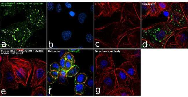
- Immunofluorescence analysis of Phospho-HER-2 / ErbB2 pTyr1221 + pTyr1222 was performed using 90% confluent log phase SK-BR-3 cells treated with 200 ng/mL of EGF for 10 minutes. The cells were fixed with 4% paraformaldehyde for 10 minutes, permeabilized with 0.1% Triton™ X-100 for 15 minutes, and blocked with 1% BSA for 1 hour at room temperature. The cells were labeled with Phospho-HER-2 / ErbB2 pTyr1221 + pTyr1222 Rabbit Polyclonal Antibody (Product # PA5-37565) at 5 µg in 0.1% BSA and incubated overnight at 4 degree Celsius and then labelled with Goat anti-Rabbit IgG (H+L) Superclonal™ Secondary Antibody, Alexa Fluor® 488 conjugate (Product # A27034) at a dilution of 1:2000 for 45 minutes at room temperature (Panel a: green). Nuclei (Panel b: blue) were stained with SlowFade® Gold Antifade Mountant with DAPI (Product # S36938). F-actin (Panel c: red) was stained with Rhodamine Phalloidin (Product # R415, 1:100). Panel d represents the merged image showing membrane and cytoplasmic localization. Panel e represents cells treated with antagonist, Afatinib (1µM for 6hrs) followed by EGF (200 ng/mL for 10 minutes), showing no signal. Panel f shows untreated cells with membrane staining. Panel g represents control cells with no primary antibody to assess background. The images were captured at 60X magnification.
- Submitted by
- Invitrogen Antibodies (provider)
- Main image

- Experimental details
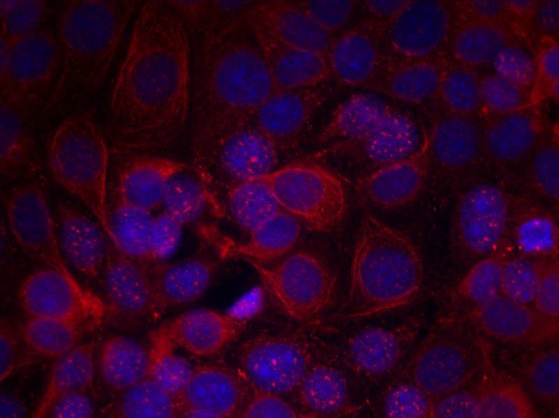
- Immunocytochemical analysis of Phospho-ErbB2 (HER-2) (Tyr1221, Tyr1222) in methanol-fixed MCF cells, using Phospho-ErbB2 (HER-2) (Tyr1221, Tyr1222) Polyclonal Antibody (Product # PA5-37565).