 Explore
 Validate
 Learn
Learn Western blot
 Immunohistochemistry
Antibody data
- Antibody Data
- Antigen structure
- References [47]
- Comments [0]
- Validations
- Western blot [2]
- Immunocytochemistry [1]
- Other assay [18]
Submit
Validation data
Reference
Comment
Report error
- Product number
- MA1-35720 - Provider product page

- Provider
- Invitrogen Antibodies
- Product name
- ErbB2 (HER-2) Monoclonal Antibody (CB11)
- Antibody type
- Monoclonal
- Antigen
- Other
- Reactivity
- Human, Mouse, Rat
- Host
- Mouse
- Isotype
- IgG
- Antibody clone number
- CB11
- Vial size
- 1 mL
- Concentration
- Conc. Not Determined
- Storage
- 4° C
Submitted references MAL2 mediates the formation of stable HER2 signaling complexes within lipid raft-rich membrane protrusions in breast cancer cells.
HER2-Mediated Internalization of Cytotoxic Agents in ERBB2 Amplified or Mutant Lung Cancers.
Hypoxia Attenuates Trastuzumab Uptake and Trastuzumab-Emtansine (T-DM1) Cytotoxicity through Redistribution of Phosphorylated Caveolin-1.
Inhibition of ezrin causes PKCα-mediated internalization of erbb2/HER2 tyrosine kinase in breast cancer cells.
HER2 signaling regulates HER2 localization and membrane retention.
The scaffolding protein NHERF1 regulates the stability and activity of the tyrosine kinase HER2.
St Gallen molecular subtypes in feline mammary carcinoma and paired metastases-disease progression and clinical implications from a 3-year follow-up study.
Heat shock protein 27 and gross cystic disease fluid protein 15 play critical roles in molecular apocrine breast cancer.
PMCA2 regulates HER2 protein kinase localization and signaling and promotes HER2-mediated breast cancer.
Fibroblastic reticular cell tumor of the breast: A case report and review of the literature.
Serum HER2 levels are increased in cats with mammary carcinomas and predict tissue HER2 status.
Synergy of leptin/STAT3 with HER2 receptor induces tamoxifen resistance in breast cancer cells through regulation of apoptosis-related genes.
Tissue biomarkers in prognostication of serous ovarian cancer following neoadjuvant chemotherapy.
Contrast enhanced computed tomography is indicative for angiogenesis pattern and display prognostic significance in breast cancer.
Ki-67 as a predictor of response to neoadjuvant chemotherapy in breast cancer patients.
Assessment of Her-2/neu status using immunocytochemistry and fluorescence in situ hybridization on fine-needle aspiration cytology smears: experience from a tertiary care centre in India.
Primary acinic cell carcinoma of the breast: a case report and review of the literature.
Prognostic and predictive significance of MYC and KRAS alterations in breast cancer from women treated with neoadjuvant chemotherapy.
An associated classification of triple negative breast cancer: the risk of relapse and the response to chemotherapy.
Are breast cancer molecular classes predictive of survival in patients with long follow-up?
Tumor-infiltrating lymphocytes, tumor characteristics, and recurrence in patients with early breast cancer.
MYC overexpression and poor prognosis in sporadic breast cancer with BRCA1 deficiency.
Lapatinib and 17AAG reduce 89Zr-trastuzumab-F(ab')2 uptake in SKBR3 tumor xenografts.
Plasma human mammaglobin mRNA associated with poor outcome in patients with breast cancer.
Epithelial-mesenchymal transition in breast cancer correlates with high histological grade and triple-negative phenotype.
Investigation of immunohistochemical ERα, ERβ and ERβcx expressions in normal and neoplastic breast tissues.
Intraoperative near-infrared fluorescence tumor imaging with vascular endothelial growth factor and human epidermal growth factor receptor 2 targeting antibodies.
Immunohistochemical COX-2 overexpression correlates with HER-2/neu overexpression in invasive breast carcinomas: a pilot study.
Survivin regulation by HER2 through NF-κB and c-myc in irradiated breast cancer cells.
The quantitative detection of total HER2 load by quantum dots and the identification of a new subtype of breast cancer with different 5-year prognosis.
Trastuzumab induced in vivo tissue remodelling associated in vitro with inhibition of the active forms of AKT and PTEN and RhoB induction in an ovarian carcinoma model.
Morphological and immunophenotypic features of primary and metastatic giant cell tumour of bone.
Dendritic cell sarcomas/tumours of the breast: report of two cases.
Basal-HER2 phenotype shows poorer survival than basal-like phenotype in hormone receptor-negative invasive breast cancers.
Expression of c-kit proto-oncogene product in breast cancer tissues.
Expression of c-kit proto-oncogene product in breast cancer tissues.
Epidermal growth factor ligand/receptor loop and downstream signaling activation pattern in completely resected nonsmall cell lung cancer.
Tumor cell expression of HLA-DM associates with a Th1 profile and predicts improved survival in breast carcinoma patients.
Prognostic and predictive value of c-erbB2 overexpression in osteogenic sarcoma.
Epidermal growth factor receptor (EGFR) expression in childhood brain tumors.
Selective inhibition of HER2 inhibits AKT signal transduction and prolongs disease-free survival in a micrometastasis model of ovarian carcinoma.
erbB-2 (HER-2) and breast cancer progression.
Intraepidermal cells of Paget's carcinoma of the breast can be genetically different from those of the underlying carcinoma.
Intraepidermal cells of Paget's carcinoma of the breast can be genetically different from those of the underlying carcinoma.
Fine-needle aspiration cytology findings of an uncommon micropapillary variant of pure mucinous carcinoma of the breast: review of patients over an 8-year period.
Toker cells are probably precursors of Paget cell carcinoma: a morphological and ultrastructural description.
Expression of erbB-4/HER-4 growth factor receptor isoforms in ovarian cancer.
Jeong J, Shin JH, Li W, Hong JY, Lim J, Hwang JY, Chung JJ, Yan Q, Liu Y, Choi J, Wysolmerski J
Cell reports 2021 Dec 28;37(13):110160
Cell reports 2021 Dec 28;37(13):110160
HER2-Mediated Internalization of Cytotoxic Agents in ERBB2 Amplified or Mutant Lung Cancers.
Li BT, Michelini F, Misale S, Cocco E, Baldino L, Cai Y, Shifman S, Tu HY, Myers ML, Xu C, Mattar M, Khodos I, Little M, Qeriqi B, Weitsman G, Wilhem CJ, Lalani AS, Diala I, Freedman RA, Lin NU, Solit DB, Berger MF, Barber PR, Ng T, Offin M, Isbell JM, Jones DR, Yu HA, Thyparambil S, Liao WL, Bhalkikar A, Cecchi F, Hyman DM, Lewis JS, Buonocore DJ, Ho AL, Makker V, Reis-Filho JS, Razavi P, Arcila ME, Kris MG, Poirier JT, Shen R, Tsurutani J, Ulaner GA, de Stanchina E, Rosen N, Rudin CM, Scaltriti M
Cancer discovery 2020 May;10(5):674-687
Cancer discovery 2020 May;10(5):674-687
Hypoxia Attenuates Trastuzumab Uptake and Trastuzumab-Emtansine (T-DM1) Cytotoxicity through Redistribution of Phosphorylated Caveolin-1.
Indira Chandran V, Månsson AS, Barbachowska M, Cerezo-Magaña M, Nodin B, Joshi B, Koppada N, Saad OM, Gluz O, Isaksson K, Borgquist S, Jirström K, Nabi IR, Jernström H, Belting M
Molecular cancer research : MCR 2020 Apr;18(4):644-656
Molecular cancer research : MCR 2020 Apr;18(4):644-656
Inhibition of ezrin causes PKCα-mediated internalization of erbb2/HER2 tyrosine kinase in breast cancer cells.
Jeong J, Choi J, Kim W, Dann P, Takyar F, Gefter JV, Friedman PA, Wysolmerski JJ
The Journal of biological chemistry 2019 Jan 18;294(3):887-901
The Journal of biological chemistry 2019 Jan 18;294(3):887-901
HER2 signaling regulates HER2 localization and membrane retention.
Jeong J, Kim W, Kim LK, VanHouten J, Wysolmerski JJ
PloS one 2017;12(4):e0174849
PloS one 2017;12(4):e0174849
The scaffolding protein NHERF1 regulates the stability and activity of the tyrosine kinase HER2.
Jeong J, VanHouten JN, Kim W, Dann P, Sullivan C, Choi J, Sneddon WB, Friedman PA, Wysolmerski JJ
The Journal of biological chemistry 2017 Apr 21;292(16):6555-6568
The Journal of biological chemistry 2017 Apr 21;292(16):6555-6568
St Gallen molecular subtypes in feline mammary carcinoma and paired metastases-disease progression and clinical implications from a 3-year follow-up study.
Soares M, Correia J, Peleteiro MC, Ferreira F
Tumour biology : the journal of the International Society for Oncodevelopmental Biology and Medicine 2016 Mar;37(3):4053-64
Tumour biology : the journal of the International Society for Oncodevelopmental Biology and Medicine 2016 Mar;37(3):4053-64
Heat shock protein 27 and gross cystic disease fluid protein 15 play critical roles in molecular apocrine breast cancer.
Liu X, Feng C, Liu J, Zhao L, Liu J, Zhang W, Liu N, Niu Y
Tumour biology : the journal of the International Society for Oncodevelopmental Biology and Medicine 2016 Jun;37(6):8027-36
Tumour biology : the journal of the International Society for Oncodevelopmental Biology and Medicine 2016 Jun;37(6):8027-36
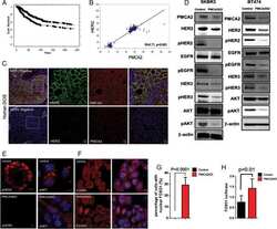
PMCA2 regulates HER2 protein kinase localization and signaling and promotes HER2-mediated breast cancer.
Jeong J, VanHouten JN, Dann P, Kim W, Sullivan C, Yu H, Liotta L, Espina V, Stern DF, Friedman PA, Wysolmerski JJ
Proceedings of the National Academy of Sciences of the United States of America 2016 Jan 19;113(3):E282-90
Proceedings of the National Academy of Sciences of the United States of America 2016 Jan 19;113(3):E282-90
Fibroblastic reticular cell tumor of the breast: A case report and review of the literature.
Li H, Shen P, Liang Y, Zhang F
Experimental and therapeutic medicine 2016 Feb;11(2):561-564
Experimental and therapeutic medicine 2016 Feb;11(2):561-564
Serum HER2 levels are increased in cats with mammary carcinomas and predict tissue HER2 status.
Soares M, Ribeiro R, Najmudin S, Gameiro A, Rodrigues R, Cardoso F, Ferreira F
Oncotarget 2016 Apr 5;7(14):17314-26
Oncotarget 2016 Apr 5;7(14):17314-26
Synergy of leptin/STAT3 with HER2 receptor induces tamoxifen resistance in breast cancer cells through regulation of apoptosis-related genes.
Papanikolaou V, Stefanou N, Dubos S, Papathanasiou I, Palianopoulou M, Valiakou V, Tsezou A
Cellular oncology (Dordrecht) 2015 Apr;38(2):155-64
Cellular oncology (Dordrecht) 2015 Apr;38(2):155-64
Tissue biomarkers in prognostication of serous ovarian cancer following neoadjuvant chemotherapy.
Khandakar B, Mathur SR, Kumar L, Kumar S, Datta Gupta S, Iyer VK, Kalaivani M
BioMed research international 2014;2014:401245
BioMed research international 2014;2014:401245
Contrast enhanced computed tomography is indicative for angiogenesis pattern and display prognostic significance in breast cancer.
Li J, Zhang Y, Zhang W, Gao Y, Jia S, Guo J
BMC cancer 2014 Sep 15;14:672
BMC cancer 2014 Sep 15;14:672
Ki-67 as a predictor of response to neoadjuvant chemotherapy in breast cancer patients.
Kim KI, Lee KH, Kim TR, Chun YS, Lee TH, Park HK
Journal of breast cancer 2014 Mar;17(1):40-6
Journal of breast cancer 2014 Mar;17(1):40-6
Assessment of Her-2/neu status using immunocytochemistry and fluorescence in situ hybridization on fine-needle aspiration cytology smears: experience from a tertiary care centre in India.
Durgapal P, Mathur SR, Kalamuddin M, Datta Gupta S, Parshad R, Julka PK, Panda SK
Diagnostic cytopathology 2014 Aug;42(8):726-31
Diagnostic cytopathology 2014 Aug;42(8):726-31
Primary acinic cell carcinoma of the breast: a case report and review of the literature.
Zhao Y, Li W, Lang R, Yang Y, Gao X, Zheng Y, Zhang C, Fu X, Fu L
International journal of surgical pathology 2014 Apr;22(2):177-81
International journal of surgical pathology 2014 Apr;22(2):177-81
Prognostic and predictive significance of MYC and KRAS alterations in breast cancer from women treated with neoadjuvant chemotherapy.
Pereira CB, Leal MF, de Souza CR, Montenegro RC, Rey JA, Carvalho AA, Assumpção PP, Khayat AS, Pinto GR, Demachki S, de Arruda Cardoso Smith M, Burbano RR
PloS one 2013;8(3):e60576
PloS one 2013;8(3):e60576
An associated classification of triple negative breast cancer: the risk of relapse and the response to chemotherapy.
Zhang J, Wang Y, Yin Q, Zhang W, Zhang T, Niu Y
International journal of clinical and experimental pathology 2013;6(7):1380-91
International journal of clinical and experimental pathology 2013;6(7):1380-91
Are breast cancer molecular classes predictive of survival in patients with long follow-up?
Pracella D, Bonin S, Barbazza R, Sapino A, Castellano I, Sulfaro S, Stanta G
Disease markers 2013;35(6):595-605
Disease markers 2013;35(6):595-605
Tumor-infiltrating lymphocytes, tumor characteristics, and recurrence in patients with early breast cancer.
Kim ST, Jeong H, Woo OH, Seo JH, Kim A, Lee ES, Shin SW, Kim YH, Kim JS, Park KH
American journal of clinical oncology 2013 Jun;36(3):224-31
American journal of clinical oncology 2013 Jun;36(3):224-31
MYC overexpression and poor prognosis in sporadic breast cancer with BRCA1 deficiency.
Ren J, Jin F, Yu Z, Zhao L, Wang L, Bai X, Zhao H, Yao W, Mi X, Wang E, Olopade OI, Wei M
Tumour biology : the journal of the International Society for Oncodevelopmental Biology and Medicine 2013 Dec;34(6):3945-58
Tumour biology : the journal of the International Society for Oncodevelopmental Biology and Medicine 2013 Dec;34(6):3945-58
Lapatinib and 17AAG reduce 89Zr-trastuzumab-F(ab')2 uptake in SKBR3 tumor xenografts.
Oude Munnink TH, de Vries EG, Vedelaar SR, Timmer-Bosscha H, Schröder CP, Brouwers AH, Lub-de Hooge MN
Molecular pharmaceutics 2012 Nov 5;9(11):2995-3002
Molecular pharmaceutics 2012 Nov 5;9(11):2995-3002
Plasma human mammaglobin mRNA associated with poor outcome in patients with breast cancer.
Lee GW, Kim JY, Koh EH, Kang D, Choi DS, Maeng KY, Lee JS
Genetics and molecular research : GMR 2012 Nov 28;11(4):4034-42
Genetics and molecular research : GMR 2012 Nov 28;11(4):4034-42
Epithelial-mesenchymal transition in breast cancer correlates with high histological grade and triple-negative phenotype.
Jeong H, Ryu YJ, An J, Lee Y, Kim A
Histopathology 2012 May;60(6B):E87-95
Histopathology 2012 May;60(6B):E87-95
Investigation of immunohistochemical ERα, ERβ and ERβcx expressions in normal and neoplastic breast tissues.
Bozkurt KK, Kapucuoğlu N
Pathology, research and practice 2012 Mar 15;208(3):133-9
Pathology, research and practice 2012 Mar 15;208(3):133-9
Intraoperative near-infrared fluorescence tumor imaging with vascular endothelial growth factor and human epidermal growth factor receptor 2 targeting antibodies.
Terwisscha van Scheltinga AG, van Dam GM, Nagengast WB, Ntziachristos V, Hollema H, Herek JL, Schröder CP, Kosterink JG, Lub-de Hoog MN, de Vries EG
Journal of nuclear medicine : official publication, Society of Nuclear Medicine 2011 Nov;52(11):1778-85
Journal of nuclear medicine : official publication, Society of Nuclear Medicine 2011 Nov;52(11):1778-85
Immunohistochemical COX-2 overexpression correlates with HER-2/neu overexpression in invasive breast carcinomas: a pilot study.
Çiriş IM, Bozkurt KK, Başpinar S, Kapucuoğlu FN
Pathology, research and practice 2011 Mar 15;207(3):182-7
Pathology, research and practice 2011 Mar 15;207(3):182-7
Survivin regulation by HER2 through NF-κB and c-myc in irradiated breast cancer cells.
Papanikolaou V, Iliopoulos D, Dimou I, Dubos S, Kappas C, Kitsiou-Tzeli S, Tsezou A
Journal of cellular and molecular medicine 2011 Jul;15(7):1542-50
Journal of cellular and molecular medicine 2011 Jul;15(7):1542-50
The quantitative detection of total HER2 load by quantum dots and the identification of a new subtype of breast cancer with different 5-year prognosis.
Chen C, Xia HS, Gong YP, Peng J, Peng CW, Hu MB, Zhu XB, Pang DW, Sun SR, Li Y
Biomaterials 2010 Nov;31(33):8818-25
Biomaterials 2010 Nov;31(33):8818-25
Trastuzumab induced in vivo tissue remodelling associated in vitro with inhibition of the active forms of AKT and PTEN and RhoB induction in an ovarian carcinoma model.
Delord JP, Quideau S, Rochaix P, Caselles O, Couderc B, Hennebelle I, Courbon F, Canal P, Allal BC
British journal of cancer 2010 Jun 29;103(1):61-72
British journal of cancer 2010 Jun 29;103(1):61-72
Morphological and immunophenotypic features of primary and metastatic giant cell tumour of bone.
Alberghini M, Kliskey K, Krenacs T, Picci P, Kindblom L, Forsyth R, Athanasou NA
Virchows Archiv : an international journal of pathology 2010 Jan;456(1):97-103
Virchows Archiv : an international journal of pathology 2010 Jan;456(1):97-103
Dendritic cell sarcomas/tumours of the breast: report of two cases.
Kapucuoglu N, Percinel S, Ventura T, Lang R, Al-Daraji W, Eusebi V
Virchows Archiv : an international journal of pathology 2009 Mar;454(3):333-9
Virchows Archiv : an international journal of pathology 2009 Mar;454(3):333-9
Basal-HER2 phenotype shows poorer survival than basal-like phenotype in hormone receptor-negative invasive breast cancers.
Liu H, Fan Q, Zhang Z, Li X, Yu H, Meng F
Human pathology 2008 Feb;39(2):167-74
Human pathology 2008 Feb;39(2):167-74
Expression of c-kit proto-oncogene product in breast cancer tissues.
Eroğlu A, Sari A
Medical oncology (Northwood, London, England) 2007;24(2):169-74
Medical oncology (Northwood, London, England) 2007;24(2):169-74
Expression of c-kit proto-oncogene product in breast cancer tissues.
Eroğlu A, Sari A
Medical oncology (Northwood, London, England) 2007;24(2):169-74
Medical oncology (Northwood, London, England) 2007;24(2):169-74
Epidermal growth factor ligand/receptor loop and downstream signaling activation pattern in completely resected nonsmall cell lung cancer.
Volante M, Saviozzi S, Rapa I, Ceppi P, Cappia S, Calogero R, Novello S, Borasio P, Papotti M, Scagliotti GV
Cancer 2007 Sep 15;110(6):1321-8
Cancer 2007 Sep 15;110(6):1321-8
Tumor cell expression of HLA-DM associates with a Th1 profile and predicts improved survival in breast carcinoma patients.
Oldford SA, Robb JD, Codner D, Gadag V, Watson PH, Drover S
International immunology 2006 Nov;18(11):1591-602
International immunology 2006 Nov;18(11):1591-602
Prognostic and predictive value of c-erbB2 overexpression in osteogenic sarcoma.
Rakesh Kumar V, Gupta N, Kakkar N, Sharma SC
Journal of cancer research and therapeutics 2006 Jan-Mar;2(1):20-3
Journal of cancer research and therapeutics 2006 Jan-Mar;2(1):20-3
Epidermal growth factor receptor (EGFR) expression in childhood brain tumors.
Bodey B, Kaiser HE, Siegel SE
In vivo (Athens, Greece) 2005 Sep-Oct;19(5):931-41
In vivo (Athens, Greece) 2005 Sep-Oct;19(5):931-41
Selective inhibition of HER2 inhibits AKT signal transduction and prolongs disease-free survival in a micrometastasis model of ovarian carcinoma.
Delord JP, Allal C, Canal M, Mery E, Rochaix P, Hennebelle I, Pradines A, Chatelut E, Bugat R, Guichard S, Canal P
Annals of oncology : official journal of the European Society for Medical Oncology 2005 Dec;16(12):1889-97
Annals of oncology : official journal of the European Society for Medical Oncology 2005 Dec;16(12):1889-97
erbB-2 (HER-2) and breast cancer progression.
Edgerton SM, Moore D 2nd, Merkel D, Thor AD
Applied immunohistochemistry & molecular morphology : AIMM 2003 Sep;11(3):214-21
Applied immunohistochemistry & molecular morphology : AIMM 2003 Sep;11(3):214-21
Intraepidermal cells of Paget's carcinoma of the breast can be genetically different from those of the underlying carcinoma.
Morandi L, Pession A, Marucci GL, Foschini MP, Pruneri G, Viale G, Eusebi V
Human pathology 2003 Dec;34(12):1321-30
Human pathology 2003 Dec;34(12):1321-30
Intraepidermal cells of Paget's carcinoma of the breast can be genetically different from those of the underlying carcinoma.
Morandi L, Pession A, Marucci GL, Foschini MP, Pruneri G, Viale G, Eusebi V
Human pathology 2003 Dec;34(12):1321-30
Human pathology 2003 Dec;34(12):1321-30
Fine-needle aspiration cytology findings of an uncommon micropapillary variant of pure mucinous carcinoma of the breast: review of patients over an 8-year period.
Ng WK
Cancer 2002 Oct 25;96(5):280-8
Cancer 2002 Oct 25;96(5):280-8
Toker cells are probably precursors of Paget cell carcinoma: a morphological and ultrastructural description.
Marucci G, Betts CM, Golouh R, Peterse JL, Foschini MP, Eusebi V
Virchows Archiv : an international journal of pathology 2002 Aug;441(2):117-23
Virchows Archiv : an international journal of pathology 2002 Aug;441(2):117-23
Expression of erbB-4/HER-4 growth factor receptor isoforms in ovarian cancer.
Gilmour LM, Macleod KG, McCaig A, Gullick WJ, Smyth JF, Langdon SP
Cancer research 2001 Mar 1;61(5):2169-76
Cancer research 2001 Mar 1;61(5):2169-76
No comments: Submit comment
Supportive validation
- Submitted by
- Invitrogen Antibodies (provider)
- Main image

- Experimental details
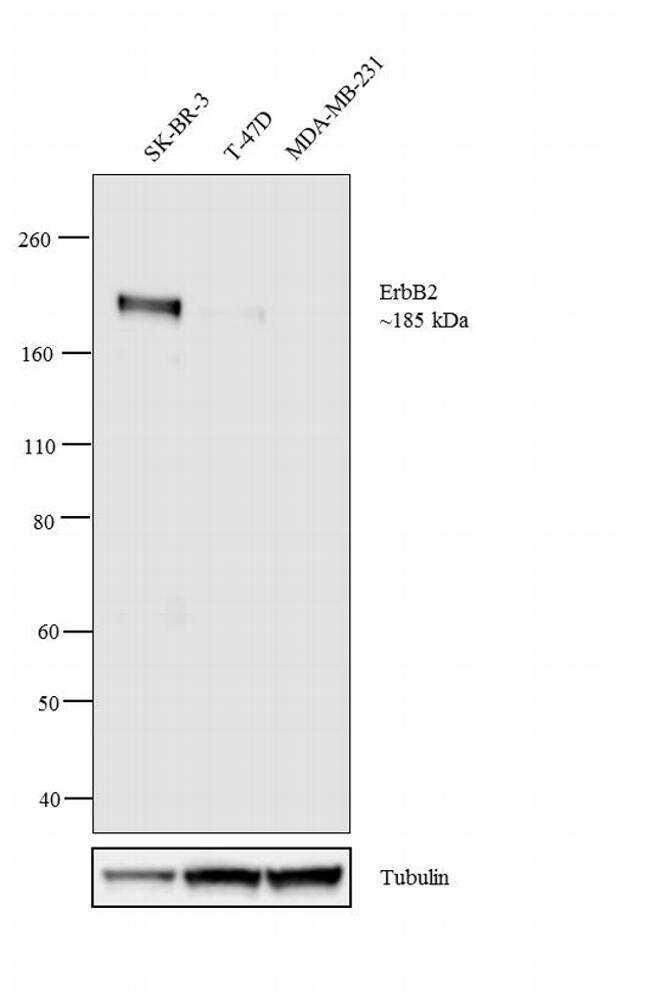
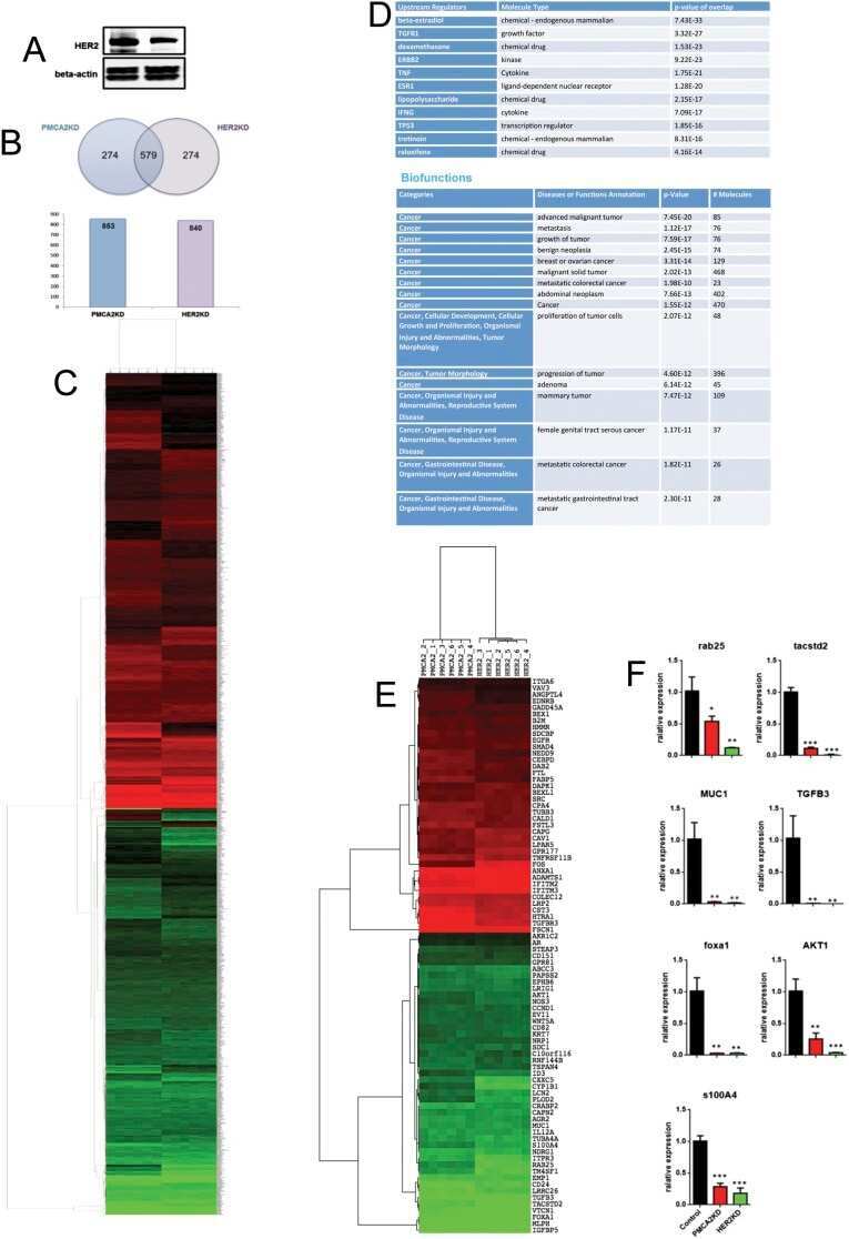
- Western blot analysis was performed on membrane enriched extracts (30 µg lysate) of SK-BR-3 (1), T-47D (2) and MDA-MB-231 (3). The blot was probed with Anti- ErbB2 Mouse Monoclonal Antibody (Product # MA1-35720, 1:1000 dilution) and detected by chemiluminescence using Goat anti-Mouse IgG (H+L) Superclonal™ Secondary Antibody, HRP conjugate (Product # A28177, 0.25 µg/mL, 1:4000 dilution). A 185 kDa band corresponding to ErbB2 was observed in SK-BR-3 and moderate in T-47D where as it was not observed in MDA-MB-231 which is an ErbB2 negative cell line. Known quantity of protein samples were electrophoresed using Novex® NuPAGE® 4-12 % Bis-Tris gel (Product # NP0321BOX), XCell SureLock™ Electrophoresis System (Product # EI0002) and Novex® Sharp Pre-Stained Protein Standard (Product # LC5800). Resolved proteins were then transferred onto a nitrocellulose membrane using the overnight wet transfer system. The membrane was probed with the relevant primary and secondary Antibody following blocking with 5 % skimmed milk. Chemiluminescent detection was performed using Pierce™ ECL Western Blotting Substrate (Product # 32106).
- Submitted by
- Invitrogen Antibodies (provider)
- Main image

- Experimental details
- Western blot analysis was performed on membrane enriched extracts (30 µg lysate) of SK-BR-3 (1), T-47D (2) and MDA-MB-231 (3). The blot was probed with Anti- ErbB2 Mouse Monoclonal Antibody (Product # MA1-35720, 1:1000 dilution) and detected by chemiluminescence using Goat anti-Mouse IgG (H+L) Superclonal™ Secondary Antibody, HRP conjugate (Product # A28177, 0.25 µg/mL, 1:4000 dilution). A 185 kDa band corresponding to ErbB2 was observed in SK-BR-3 and moderate in T-47D where as it was not observed in MDA-MB-231 which is an ErbB2 negative cell line. Known quantity of protein samples were electrophoresed using Novex® NuPAGE® 4-12 % Bis-Tris gel (Product # NP0321BOX), XCell SureLock™ Electrophoresis System (Product # EI0002) and Novex® Sharp Pre-Stained Protein Standard (Product # LC5800). Resolved proteins were then transferred onto a nitrocellulose membrane using the overnight wet transfer system. The membrane was probed with the relevant primary and secondary Antibody following blocking with 5 % skimmed milk. Chemiluminescent detection was performed using Pierce™ ECL Western Blotting Substrate (Product # 32106).

Supportive validation
- Submitted by
- Invitrogen Antibodies (provider)
- Main image
- Experimental details
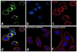
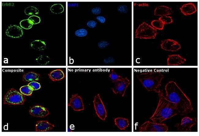
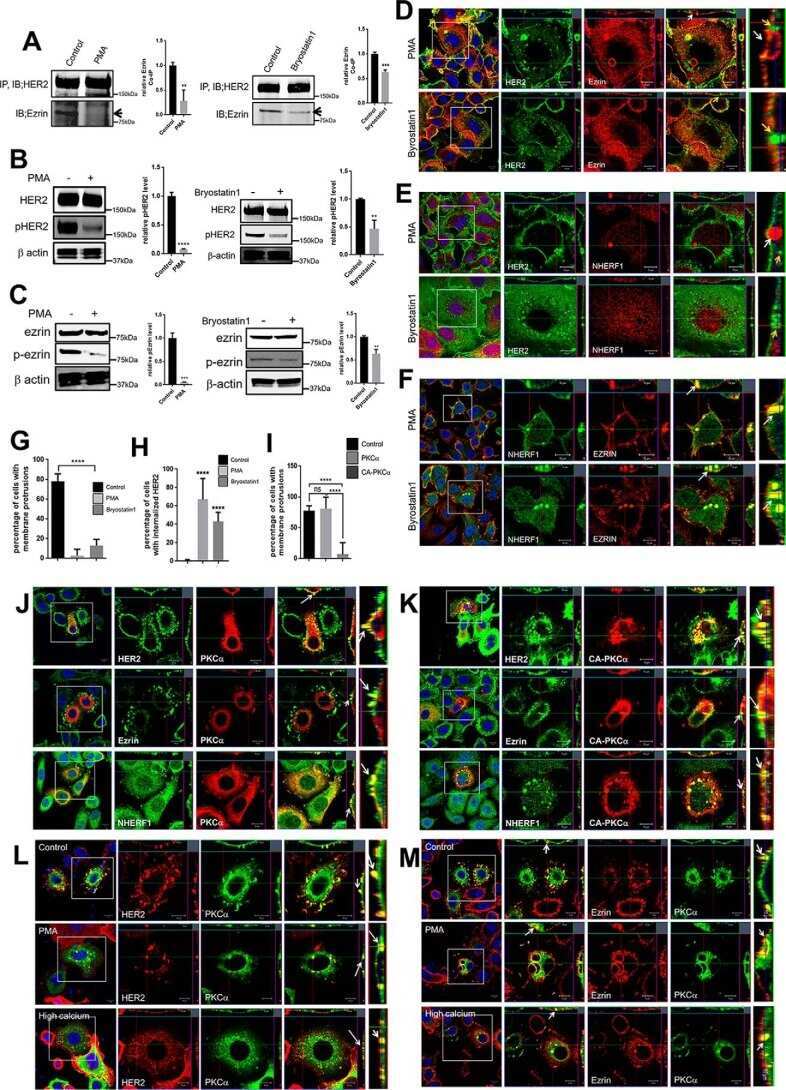
- Immunofluorescence analysis of ErbB2 was performed using 70% confluent log phase SK-BR-3 cells. The cells were fixed with 4% paraformaldehyde for 10 minutes, permeabilized with 0.1% Triton™ X-100 for 10 minutes, and blocked with 1% BSA for 1 hour at room temperature. The cells were labeled with Her2 Mouse monoclonal Antibody (Product # MA1-35720) at a dilution of 1:100 in 0.1% BSA and incubated overnight at 4 degree Celsius and then labeled with Goat anti-Mouse IgG (H+L) Superclonal™ Secondary Antibody, Alexa Fluor® 488 conjugate (Product # A28175) at a dilution of 1:2000 for 45 minutes at room temperature (Panel a: green). Nuclei (Panel b: blue) were stained with SlowFade® Gold Antifade Mountant with DAPI (Product # S36938). F-actin (Panel c: red) was stained with Rhodamine Phalloidin (Product # R415, 1:300). Panel d represents the merged image showing membranous, cytoplasmic localization. Panel f represents MDAMB-231 cells as negative controls, showing no Her2 staining. Panel e represents control cells with no primary antibody to assess background. The images were captured at 60X magnification.
Supportive validation
- Submitted by
- Invitrogen Antibodies (provider)
- Main image

- Experimental details
- NULL
- Submitted by
- Invitrogen Antibodies (provider)
- Main image
- Experimental details
- NULL

- Submitted by
- Invitrogen Antibodies (provider)
- Main image
- Experimental details
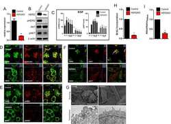
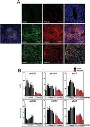
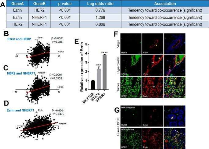
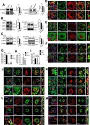
- Fig 1 Loss of HER2 prevents the formation of membrane protrusion in HER2KD-SKBR3 cells. A) Examination of relative expression of HER2 by QPCR. Bars represent mean +- SEM for 3 separate experiments. B) Immunoblot analysis from control and HER2KD-SKBR3 cells. C) Time course of pHER2 and pAKT levels in control (black bars) and HER2KD-SKBR3 (gray bars) cells in response to EGF. Cells were serum-starved for 16 hours, treated with growth factors and harvested at times listed on graphs. Each bar represents the mean +- SEM of 3 separate experiments. * represents p

- Submitted by
- Invitrogen Antibodies (provider)
- Main image
- Experimental details
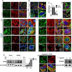
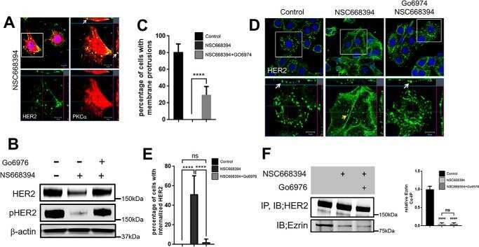
- Fig 5 HER2 signaling prevents HER2 internalization. A) Con-focal images of immunofluorescence for HER2 (green) and EGFR (red) in control (top row) and HER2KD-SKBR3 (bottom row) cells at baseline in serum-containing media. White arrow points to internalized HER2. Exposure times are longer in HER2KD cells to compensate for reduced HER2 levels. B) Con-focal images of immunofluorescence for HER2 (green) and EGFR (red) in control (top row) and HER2KD-SKBR3 (bottom row) cells exposed EGF for 2 hrs. Panels on right represent magnifications of boxed areas. White arrow indicates internalized HER2 and EGFR. C) Quantification of cell surface HER2 as detected by isolation of cell-surface biotinylated proteins. Each bar represents the mean +- SEM of 3 separate experiments in cells at baseline and after EGF treatment. Asterisk denotes p
- Submitted by
- Invitrogen Antibodies (provider)
- Main image
- Experimental details
- NULL
- Submitted by
- Invitrogen Antibodies (provider)
- Main image
- Experimental details
- NULL
- Submitted by
- Invitrogen Antibodies (provider)
- Main image
- Experimental details
- NULL

- Submitted by
- Invitrogen Antibodies (provider)
- Main image

- Experimental details
- NULL
- Submitted by
- Invitrogen Antibodies (provider)
- Main image
- Experimental details
- NULL

- Submitted by
- Invitrogen Antibodies (provider)
- Main image
- Experimental details
- NULL

- Submitted by
- Invitrogen Antibodies (provider)
- Main image
- Experimental details
- NULL
- Submitted by
- Invitrogen Antibodies (provider)
- Main image
- Experimental details
- NULL
- Submitted by
- Invitrogen Antibodies (provider)
- Main image
- Experimental details
- NULL

- Submitted by
- Invitrogen Antibodies (provider)
- Main image
- Experimental details
- NULL
- Submitted by
- Invitrogen Antibodies (provider)
- Main image
- Experimental details
- NULL
- Submitted by
- Invitrogen Antibodies (provider)
- Main image
- Experimental details
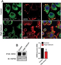
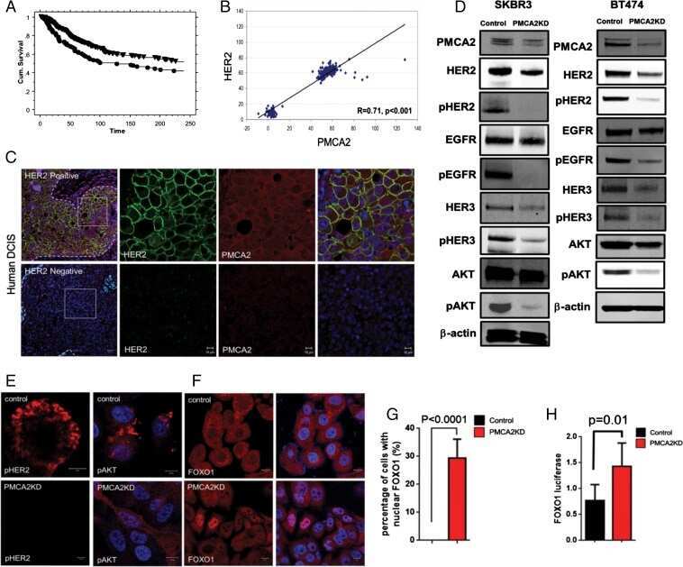
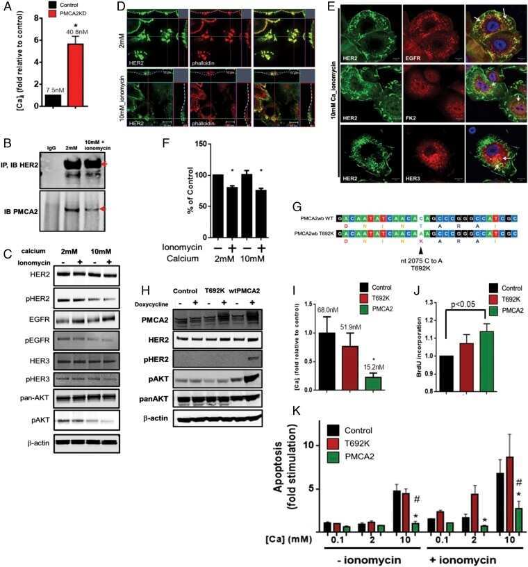
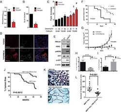
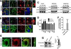
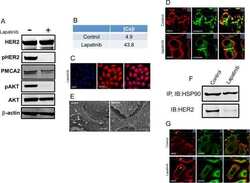
- Fig 4 Lapatinib increases intracellular calcium and inhibits membrane protrusions. A) Immunoblot showing HER2 signaling components in SKBR3 cells +- lapatinib ( 2 micromolar for 48-hours). B) Intracellular calcium measurements in control and lapatinib-treated SKBR3 cells. Numbers are mean calcium concentrations estimated by FURA2 measurements. C) Con-focal images of immunofluorescence for NFATc1 (red) and DAPI (blue) in SKBR3 cells treated with lapatinib. D) Con-focal images of immunofluorescence for HER2 (red) and phalloidin (actin, green) in control and lapatinib-treated SKBR3 cells. Insets represent Z-stack images in 2 different orientations. E) Scanning electron microscopy of control and lapatinib-treated SKBR3 cells. Arrows point to larger membrane protrusions. F) Co-IP for HER2 and HSP90 from control and lapatinib-treated SKBR3 cells. Cell lysates were immunoprecipitated with antibodies to HSP90 and blotted for HSP90 or HER2. G) Con-focal images of immunofluorescence for HER2 (red) and HSP90 (green) in control (top row) and lapatinib-treated (bottom row) SKBR3 cells. Insets represent Z-stack images in 2 different orientations and white arrows indicate internalized HER2. Scale bars in C, D and G represent 10mum. Scale bars in E represent 20muM.

- Submitted by
- Invitrogen Antibodies (provider)
- Main image
- Experimental details
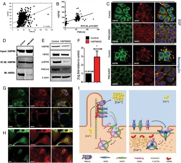
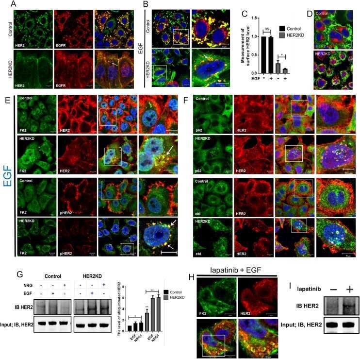
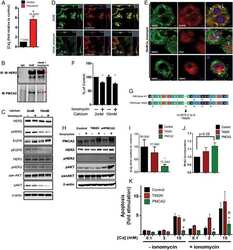
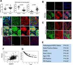
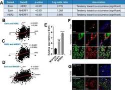
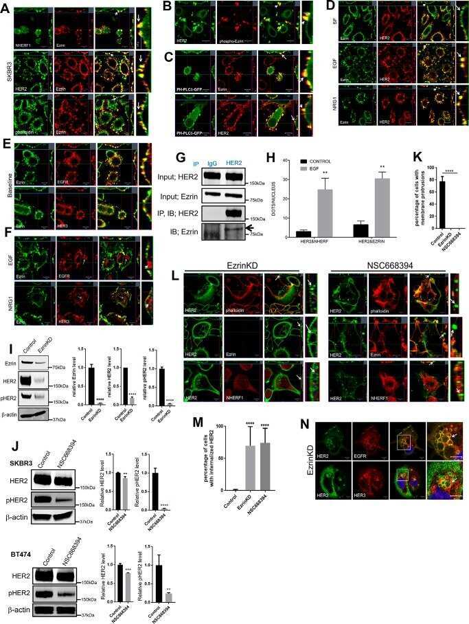
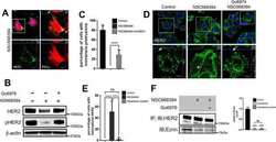
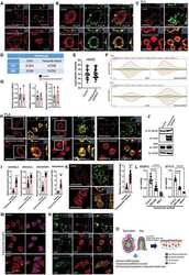
- Figure 6. Enhanced interactions between HER2 and MAL2, Ezrin, NHERF1, and PMCA2 in trastuzumab-resistant SKBR3 cells (A) Immunofluorescence staining for cholera toxin B (lipid rafts) in control and trastuzumab-resistant SKBR3 cells. Scale bars represent 10 mum. (B) Immunofluorescence staining for HER2 and MAL2 in control and trastuzumab-resistant SKBR3 cells. Scale bars represent 10 mum. (C) PLA for HER2 and MAL2 in control and trastuzumab-resistant SKBR3 cells also stained for phalloidin. Scale bars represent 10 mum. (D-F) Quantitative results from immunoprecipitation coupled with data-independent acquisition mass spectrometry (DIA-MS) in control and trastuzumab-resistant SKBR3 cells. (D) The DIA-MS Intensity (log 2 ) of HER2 and MAL2 proteins from control and trastuzumab-resistant SKBR3 cells. (E) The DIA-MS Intensity (log 2 ) for all the peptide precursor signals of HER2 in control and resistant cells. (F) The DIA-MS peak groups visualized for quantifying HER2 (VLGSGAFGTVYK) and MAL2 (VTLPAGPDILR). Peaks above and below the middle line denote the MS2 and MS1 ion traces in DIA-MS. (G) MAL2, Ezrin, and NHERF1 mRNA expression in control and trastuzumab-resistant SKBR3 cells as assessed by quantitative RT-PCR (n = 3). (H) PLA for HER2 with Ezrin (left), NHERF1 (middle), and PMCA2 (right) in control and trastuzumab-resistant SKBR3 cells also stained for phalloidin. Boxed portions are amplified at right with co-registration of PLA signal and immunofluorescence for actin (phalloi

- Submitted by
- Invitrogen Antibodies (provider)
- Main image
- Experimental details
- NULL
