 Explore
 Validate
 Learn
Learn Western blot
Antibody data
- Antibody Data
- Antigen structure
- References [0]
- Comments [0]
- Validations
- Western blot [2]
- ELISA [1]
- Immunocytochemistry [3]
- Chromatin Immunoprecipitation [2]
- Other assay [3]
Submit
Validation data
Reference
Comment
Report error
- Product number
- PA5-40100 - Provider product page

- Provider
- Invitrogen Antibodies
- Product name
- H2BK12ac Polyclonal Antibody
- Antibody type
- Polyclonal
- Antigen
- Synthetic peptide
- Reactivity
- Human
- Host
- Rabbit
- Isotype
- IgG
- Vial size
- 50 µg
- Concentration
- 0.82 mg/mL
- Storage
- -20°C or -80°C if preferred
No comments: Submit comment
Supportive validation
- Submitted by
- Invitrogen Antibodies (provider)
- Main image
- Experimental details
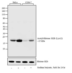
- Western blot analysis was performed on acid extracts (20 µg lysate) of HeLa (Lane 1), HeLa treated with sodium butyrate (5 nM for 24 hr) (Lane 2), COS-7 (Lane 3), and COS-7 treated with sodium butyrate (5 nM for 24 hr) (Lane 4). The blot was probed with Anti-Acetyl-Histone H2B (Lys12) polyclonal antibody (Product # PA5-40100, 1:1000 dilution) and detected by chemiluminescence using Goat anti-Rabbit IgG (H+L) Superclonal™ Secondary Antibody, HRP conjugate (Product # A27036, 0.25 µg/mL, 1:4000 dilution). A 15 kDa band corresponding to Acetyl-Histone H2B (Lys12) was observed across the cell lines tested and enhanced upon treatment.

- Submitted by
- Invitrogen Antibodies (provider)
- Main image
- Experimental details
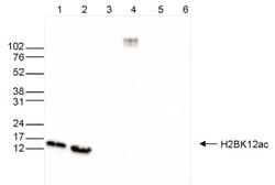
- Western Blot was performed on whole cell (25 µg, lane 1) and histone extracts (15 µg, lane 2) from HeLa cells, and on 1 µg of recombinant histone H2A, H2B, H3 and H4 (lane 3, 4, 5 and 6, respectively) using a Acetyl-Histone H2B (Lys12) polyclonal antibody (Product # PA5-40100) at a dilution of 1:1,000 in TBS-Tween containing 5% skimmed milk. The marker (kDa) is shown on the left.

Supportive validation
- Submitted by
- Invitrogen Antibodies (provider)
- Main image
- Experimental details
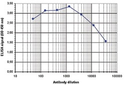
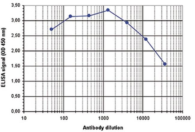
- To determine the titer of the antibody, an ELISA was performed using a serial dilution of the anti-H2BK12ac antibody (Product # PA5-40100) in antigen coated wells. The antigen used was a peptide containing the histone modification of interest. By plotting the absorbance against the antibody dilution, the titer of the antibody was estimated to be 1:38,200.
Supportive validation
- Submitted by
- Invitrogen Antibodies (provider)
- Main image

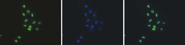
- Experimental details
- HeLa cells were stained with the anti-H2BK12ac antibody (Product # PA5-40100) and with DAPI. Cells were fixed with 4% formaldehyde for 10’ and blocked with PBS/TX-100 containing 5% normal goat serum and 1% BSA. The cells were immunofluorescently labeled with the H2BK12ac antibody (left) diluted 1:500 in blocking solution followed by an anti-rabbit antibody conjugated to Alexa488. The middle panel shows staining of the nuclei with DAPI. A merge of the two stainings is shown on the right.
- Submitted by
- Invitrogen Antibodies (provider)
- Main image

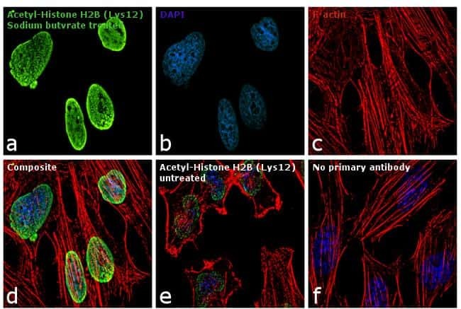
- Experimental details
- Immunofluorescence analysis of Acetyl-Histone H2B (Lys12) was performed using 70% confluent log phase HeLa cells treated with sodium butyrate. The cells were fixed with 4% paraformaldehyde for 10 minutes, permeabilized with 0.1% Triton™ X-100 for 10 minutes, and blocked with 1% BSA for 1 hour at room temperature. The cells were labeled with Acetyl-Histone H2B (Lys12) Rabbit Polyclonal Antibody (Product # PA5-40100) at 1:100 dilution in 0.1% BSA, incubated overnight at 4 degree Celsius and then labeled with Goat anti-Rabbit IgG (H+L) Superclonal™ Secondary Antibody, Alexa Fluor® 488 conjugate (Product # A27034) at a dilution of 1:2000 for 45 minutes at room temperature (Panel a: green). Nuclei (Panel b: blue) were stained with SlowFade® Gold Antifade Mountant with DAPI (Product # S36938). F-actin (Panel c: red) was stained with Rhodamine Phalloidin (Product # R415, 1:300). Panel d represents the merged image showing nuclear localization. Panel e represents the untreated cells with relatively lower expression of Acetyl-Histone H2B (Lys12) polyclonal Antibody. Panel f shows control cells with no primary antibody to assess background. The images were captured at 60X magnification.
- Submitted by
- Invitrogen Antibodies (provider)
- Main image

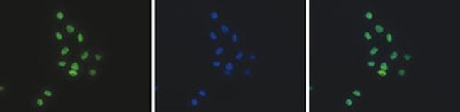
- Experimental details
- HeLa cells were stained with the anti-H2BK12ac antibody (Product # PA5-40100) and with DAPI. Cells were fixed with 4% formaldehyde for 10’ and blocked with PBS/TX-100 containing 5% normal goat serum and 1% BSA. The cells were immunofluorescently labeled with the H2BK12ac antibody (left) diluted 1:500 in blocking solution followed by an anti-rabbit antibody conjugated to Alexa488. The middle panel shows staining of the nuclei with DAPI. A merge of the two stainings is shown on the right.
Supportive validation
- Submitted by
- Invitrogen Antibodies (provider)
- Main image
- Experimental details
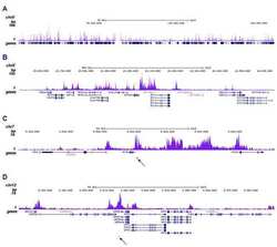
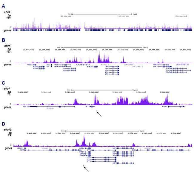
- ChIP was performed on sheared chromatin from 1.5 million HeLaS3 cells using 0.5 µg of Acetyl-Histone H2B (Lys12) polyclonal antibody (Product # PA5-40100). The IP'd DNA was subsequently analyzed on a HiSeq. The 51 bp tags were aligned to the human genome using the BWA algorithm. Figure 2 shows the enrichment along the complete sequence and a 1 Mb region of the X-chromosome (fig 2A and B) and in genomic regions of chromosome 7, surrounding the ACTB gene, and of chromosome 12, surrounding the GAPDH gene (fig 2C and D). The position of the amplicon used for ChIP-qPCR is indicated by an arrow.
- Submitted by
- Invitrogen Antibodies (provider)
- Main image
- Experimental details
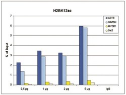
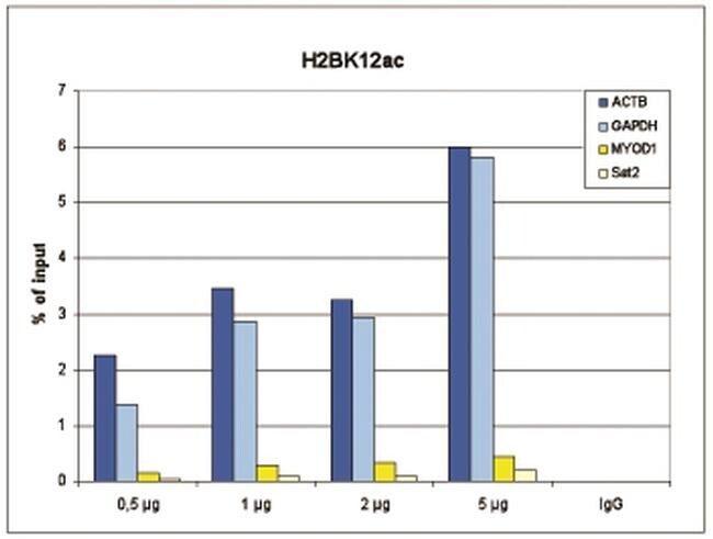
- ChIP assays were performed on human HeLa cells using a Acetyl-Histone H2B (Lys12) polyclonal antibody (Product # PA5-40100) and optimized PCR primer sets for qPCR. ChIP was performed with sheared chromatin from 1.5 million cells. A titration of the antibody consisting of 0.5, 1, 2 and, 5 µg per ChIP experiment was analyzed. IgG (1 µg/IP) was used as negative IP control. QPCR was performed with primers for a region approximately 1 kb upstream of the GAPDH and ACTB promoters, used as positive controls, and for the coding region of the inactive MYOD1 gene and the Sat2 satellite repeat, used as negative controls. Figure 1 shows the recovery, expressed as a % of input (the relative amount of immunoprecipitated DNA compared to input DNA after qPCR analysis).
Supportive validation
- Submitted by
- Invitrogen Antibodies (provider)
- Main image

- Experimental details
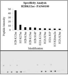
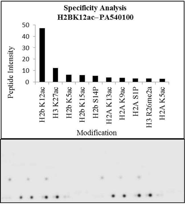
- Antibody specificity for modified targets can be established using peptide arrays by quantifying detection of the target protein along with closely related proteins. Peptide array of Histone H2BK12ac using Anti-Acetyl-Histone H2B (Lys12) Antibody: An array of the specific peptide and other relevant peptides when tested using Anti-Acetyl-Histone H2B (Lys12) Polyclonal Antibody (Product # PA5-40100), showed that the Histone H2BK12ac modification was specifically recognized by the antibody.

- Submitted by
- Invitrogen Antibodies (provider)
- Main image
- Experimental details
- Antibody specificity for modified targets can be established using peptide arrays by quantifying detection of the target protein along with closely related proteins. Peptide array of Histone H2BK12ac using Anti-Acetyl-Histone H2B (Lys12) Antibody: An array of the specific peptide and other relevant peptides when tested using Anti-Acetyl-Histone H2B (Lys12) Polyclonal Antibody (Product # PA5-40100), showed that the Histone H2BK12ac modification was specifically recognized by the antibody.
- Submitted by
- Invitrogen Antibodies (provider)
- Main image
- Experimental details
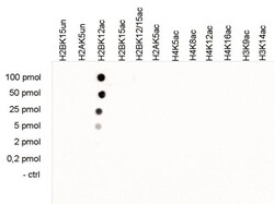
- A Dot Blot analysis was performed to test the cross reactivity of Acetyl-Histone H2B (Lys12) polyclonal antibody (Product # PA5-40100) with peptides containing other histone modifications and the unmodified H2B. One hundred to 0.2 pmol of the respective peptides were spotted on a membrane. The antibody was used at a dilution of 1:5,000. Figure 4 shows a high specificity of the antibody for the modification of interest.
