Explore
 Validate
 Learn
Learn Western blot
 ELISA
ELISAAntibody data
- Antibody Data
- Antigen structure
- References [2]
- Comments [0]
- Validations
- Western blot [1]
- Immunocytochemistry [1]
- Immunohistochemistry [1]
- Flow cytometry [1]
Submit
Validation data
Reference
Comment
Report error
- Product number
- ABIN954456 - Provider product page

- Provider
- antibodies-online
- Product name
- anti-Arginyl-tRNA Synthetase (RARS) (C-Term) antibody
- Antibody type
- Polyclonal
- Antigen
- Other
- Reactivity
- Human
- Host
- Rabbit
- Vial size
- 0.1 mg
Submitted references Proteasomes and RARS modulate AIMP1/EMAP II secretion in human cancer cell lines.
Exploring proteomes and analyzing protein processing by mass spectrometric identification of sorted N-terminal peptides.
Bottoni A, Vignali C, Piccin D, Tagliati F, Luchin A, Zatelli MC, Uberti EC
Journal of cellular physiology 2007 Aug;212(2):293-7
Journal of cellular physiology 2007 Aug;212(2):293-7
Exploring proteomes and analyzing protein processing by mass spectrometric identification of sorted N-terminal peptides.
Gevaert K, Goethals M, Martens L, Van Damme J, Staes A, Thomas GR, Vandekerckhove J
Nature biotechnology 2003 May;21(5):566-9
Nature biotechnology 2003 May;21(5):566-9
No comments: Submit comment
Supportive validation
- Submitted by
- antibodies-online (provider)
- Main image
- Experimental details
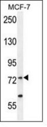
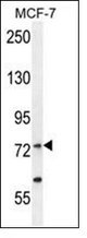
- Western blot analysis of RARS Antibody (C-term) Cat.-No AP53582PU-N in MCF-7 cell line lysates (35ug/lane). This demonstrates the RARS antibody detected the RARS protein (arrow).
Supportive validation
- Submitted by
- antibodies-online (provider)
- Main image

- Experimental details
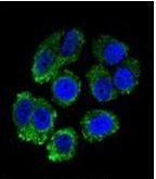
- Confocal immunofluorescent analysis of RARS Antibody (C-term) Cat.-No AP53582PU-N with MCF-7 cell followed by Alexa Fluor 488-conjugated Goat anti-Rabbit lgG (green). DAPI was used to stain the cell nuclear (blue).
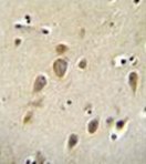
Supportive validation
- Submitted by
- antibodies-online (provider)
- Main image
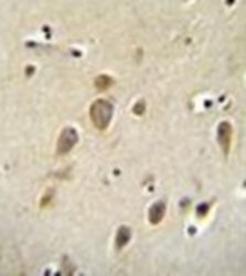
- Experimental details
- Formalin fixed, paraffin embedded human brain stained with RARS Antibody (C-term) Cat.-No AP53582PU-N followed by peroxidase conjugation of the secondary antibody and DAB staining.
Supportive validation
- Submitted by
- antibodies-online (provider)
- Main image
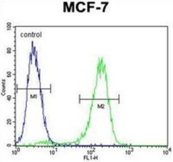
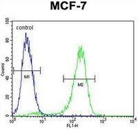
- Experimental details
- Flow cytometric analysis of MCF-7 cells using RARS Antibody (C-term) Cat.-No AP53582PU-N (right histogram) compared to a negative control cell (left histogram). FITC-conjugated goat-anti-rabbit secondary antibodies were used for the analysis.