Explore
 Validate
 Learn
Learn Western blot
 ELISA
ELISAAntibody data
- Antibody Data
- Antigen structure
- References [3]
- Comments [0]
- Validations
- Western blot [1]
- Flow cytometry [1]
Submit
Validation data
Reference
Comment
Report error
- Product number
- ABIN238332 - Provider product page

- Provider
- antibodies-online
- Proper citation
- Antibodies-Online Cat#ABIN238332, RRID:AB_10952471
- Product name
- anti-Acid Phosphatase, Prostate (ACPP) antibody
- Antibody type
- Monoclonal
- Antigen
- genetic immunisation with cDNA encoding human PAP
- Description
- Protein G
- Reactivity
- Human
- Isotype
- IgG
- Antibody clone number
- LT-3D1
- Vial size
- 100 μg
- Concentration
- 2 mg/mL
- Storage
- short term: 2°C - 8°C, long term: -20°C
- Handling
- Avoid repeated freezing and thawing.
Submitted references Cellular prostatic acid phosphatase: a protein tyrosine phosphatase involved in androgen-independent proliferation of prostate cancer.
Human prostatic acid phosphatase: selected properties and practical applications.
Prostate-specific antigen and prostatic acid phosphatase: biomolecular and physiologic characteristics.
Veeramani S, Yuan TC, Chen SJ, Lin FF, Petersen JE, Shaheduzzaman S, Srivastava S, MacDonald RG, Lin MF
Endocrine-related cancer 2005 Dec;12(4):805-22
Endocrine-related cancer 2005 Dec;12(4):805-22
Human prostatic acid phosphatase: selected properties and practical applications.
Ostrowski WS, Kuciel R
Clinica chimica acta; international journal of clinical chemistry 1994 May;226(2):121-9
Clinica chimica acta; international journal of clinical chemistry 1994 May;226(2):121-9
Prostate-specific antigen and prostatic acid phosphatase: biomolecular and physiologic characteristics.
Bilhartz DL, Tindall DJ, Oesterling JE
Urology 1991 Aug;38(2):95-102
Urology 1991 Aug;38(2):95-102
No comments: Submit comment
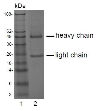
Supportive validation
- Submitted by
- antibodies-online (provider)
- Main image
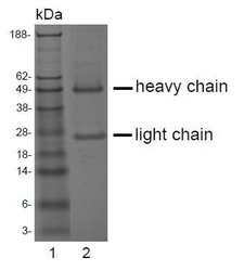
- Experimental details
- SDS-PAGE analysis of purified LT-3D1 monoclonal antibody. Lane 1: molecular weight marker, Lane 2: 2 ?g of purified LT-3D1 antibody. Proteins were separated by SDS-PAGE and stained with RAPID StainTM Reagent.
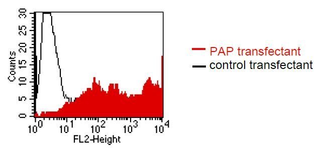
Supportive validation
- Submitted by
- antibodies-online (provider)
- Main image
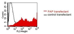
- Experimental details
- FACS analysis of BOSC23 cells using LT-3D1. BOSC23 cells were transiently transfected with an expres-sion vector encoding either PAP (red curve) or an irrelevant protein (control transfectant: black curve). Binding of LT-3D1 was detected with a PE-conjugated secondary antibody. A positive signal was obtained only with PAP transfected cells.