Explore
 Validate
 Learn
Learn Western blot
Antibody data
- Antibody Data
- Antigen structure
- References [0]
- Comments [0]
- Validations
- Western blot [2]
Submit
Validation data
Reference
Comment
Report error
- Product number
- PA1-331B - Provider product page

- Provider
- Invitrogen Antibodies
- Product name
- LXR alpha/beta Polyclonal Antibody
- Antibody type
- Polyclonal
- Antigen
- Synthetic peptide
- Description
- PA1-331B detects recombinant human liver X receptor (LXR). PA1-331B has been successfully used in Western blot procedures. By Western blot, this antibody detects an ~64 kDa and ~80 kDa protein representing recombinant human LXR alpha and beta. The PA1-331B immunogen is a synthetic peptide corresponding to residues C L(432) R L Q D K K L P P L L S E(445) of human LXR alpha. This sequence also corresponds to amino acids 442-455 of the human LXR beta. PA1-331B immunizing peptide (Cat. # PEP-115) is available for use in neutralization and control experiments.
- Reactivity
- Human
- Host
- Rabbit
- Isotype
- IgG
- Vial size
- 200 µg
- Concentration
- 1 mg/mL
- Storage
- -20° C, Avoid Freeze/Thaw Cycles
No comments: Submit comment
Supportive validation
- Submitted by
- Invitrogen Antibodies (provider)
- Main image
- Experimental details
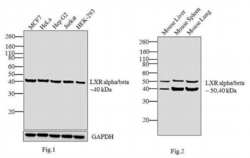
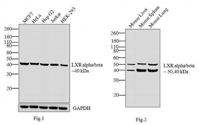
- Fig.1) Western blot analysis was performed on nuclear enriched extracts (30 µglysate) of MCF7 (Lane 1), HeLa (Lane 2), Hep G2 (Lane 3), Jurkat (Lane 4) and HEK-293 (Lane 5). Fig2.) Likewise, western blot analysis was performed on tissue extracts (30 µglysate) of Mouse Liver, Mouse Spleen and Mouse Lung. The blots were probed with LXR alpha/beta Rabbit polyclonal Antibody (Product # PA1-331B, 2 µg/mL) and detected by chemiluminescence using Goat anti-Rabbit IgG (H+L) Superclonal™ Secondary Antibody, HRP conjugate (Product # A27036, 0.4 µg/mL, 1:2500 dilution). A 40 kDa band corresponding to LXR beta was observed across the cell lines tested. Apart from this in Fig. 2, 50 kDa corresponding to LXR alpha form is also observed across all tissue extracts tested. Known quantity of protein samples were electrophoresed using Novex® NuPAGE® 4-12 % Bis-Tris gel (Product # NP0322BOX), XCell SureLock™ Electrophoresis System (Product # EI0002) and Novex® Sharp Pre-Stained Protein Standard (Product # LC5800). Resolved proteins were then transferred onto a nitrocellulose membrane with iBlot® 2 Dry Blotting System (Product # IB21001). The membrane was probed with the relevant primary and secondary Antibody following blocking with 5% skimmed milk. Chemiluminescent detection was performed using Pierce™ ECL Western Blotting Substrate (Product # 32106).
- Submitted by
- Invitrogen Antibodies (provider)
- Main image
- Experimental details
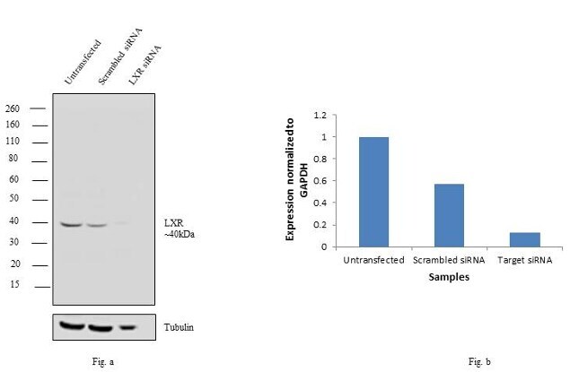
- Knockdown of LXR alpha/beta was achieved by transfecting HeLa cells with LXR alpha/beta specific siRNAs (Silencer® select Product # s14684, s19568). Western blot analysis (Fig a) was performed using whole cell extracts from LXR alpha/beta knock down cells (lane 3), non-specific scrambled siRNA transfected cells (lane 2) and untransfected cells (lane 1). The blots were probed with Anti- LXR alpha/beta Rabbit polyclonal Antibody (Product # PA1-331B, 1µg/mL) and Goat anti-Rabbit IgG (H+L) Superclonal™ Secondary Antibody, HRP conjugate (Product # A27036, 0.25 µg/mL, 1:4000 dilution). Densitometric analysis of this western blot is shown in histogram (Fig b). Decrease in signal upon siRNA mediated knock down confirms that antibody is specific to LXR alpha/beta.