Explore
 Validate
 Learn
Learn Immunocytochemistry
Antibody data
- Antibody Data
- Antigen structure
- References [19]
- Comments [0]
- Validations
- Immunocytochemistry [1]
- Other assay [10]
Submit
Validation data
Reference
Comment
Report error
- Product number
- 41-9003-80 - Provider product page

- Provider
- Invitrogen Antibodies
- Product name
- Pan Cytokeratin Monoclonal Antibody (AE1/AE3), eFluor™ 570, eBioscience™
- Antibody type
- Monoclonal
- Antigen
- Other
- Description
- Description: The monoclonal antibodies AE1 and AE3 recognize many of the acidic and basic cytokeratin family members. Cytokeratins are intermediate filament proteins comprising one component of the cytoskeleton. There are two large families of cytokeratins, acidic and basic, but all contain the same basic domains (i.e. an alpha-helical core with an N- and C-terminal domain). The proteins are expressed in epithelial cells, but are developmentally regulated. Many tumors also express these proteins and their expression can help identify the origin of a neoplasm.
- Antibody clone number
- AE1/AE3
- Concentration
- 0.2 mg/mL
Submitted references Spine Metastases in Immunocompromised Mice after Intracardiac Injection of MDA-MB-231-SCP2 Breast Cancer Cells.
Application of circulating tumour cells to predict response to treatment in head and neck cancer.
The Extracellular Small Leucine-Rich Proteoglycan Biglycan Is a Key Player in Gastric Cancer Aggressiveness.
Molecular Features of Cancers Exhibiting Exceptional Responses to Treatment.
Prognostic impact of CD73 expression and its relationship to PD-L1 in patients with radically treated pancreatic cancer.
Cysteine-Rich Angiogenic Inducer 61: Pro-Survival Function and Role as a Biomarker for Disseminating Breast Cancer Cells.
Effective Reconstruction of Functional Urethra Promoted With ICG-001 Delivery Using Core-Shell Collagen/Poly(Llactide-co-caprolactone) [P(LLA-CL)] Nanoyarn-Based Scaffold: A Study in Dog Model.
Characterization of circulating breast cancer cells with tumorigenic and metastatic capacity.
PD-L1 Expression on Circulating Tumour Cells May Be Predictive of Response to Regorafenib in Patients Diagnosed with Chemorefractory Metastatic Colorectal Cancer.
Microsieves for the detection of circulating tumor cells in leukapheresis product in non-small cell lung cancer patients.
Clonality of circulating tumor cells in breast cancer brain metastasis patients.
Screening Circulating Tumor Cells as a Noninvasive Cancer Test in 3388 Individuals from High-Risk Groups (ICELLATE2).
Scanning Electron Microscopy of Circulating Tumor Cells and Tumor-Derived Extracellular Vesicles.
VyCAP's puncher technology for single cell identification, isolation, and analysis.
Neither epithelial nor mesenchymal circulating tumor cells isolated from breast cancer patients are tumorigenic in NOD-scid Il2rg(null) mice.
EGFR-GRB2 Protein Colocalization Is a Prognostic Factor Unrelated to Overall EGFR Expression or EGFR Mutation in Lung Adenocarcinoma.
Identification of a Hematopoietic Cell Dedifferentiation-Inducing Factor.
Flow cytometric determination of stem/progenitor content in epithelial tissues: an example from nonsmall lung cancer and normal lung.
KIT (CD117) expression in a subset of non-small cell lung carcinoma (NSCLC) patients.
Brylka L, Jähn-Rickert K, Baranowsky A, Neven M, Horn M, Yorgan T, Wikman H, Werner S, Lübke A, Amling M, Busse B, Pantel K, Schinke T
Cancers 2022 Jan 22;14(3)
Cancers 2022 Jan 22;14(3)
Application of circulating tumour cells to predict response to treatment in head and neck cancer.
Zhang X, Ekanayake Weeramange C, Hughes BGM, Vasani S, Liu ZY, Warkiani ME, Hartel G, Ladwa R, Thiery JP, Kenny L, Punyadeera C
Cellular oncology (Dordrecht) 2022 Aug;45(4):543-555
Cellular oncology (Dordrecht) 2022 Aug;45(4):543-555
The Extracellular Small Leucine-Rich Proteoglycan Biglycan Is a Key Player in Gastric Cancer Aggressiveness.
Pinto F, Santos-Ferreira L, Pinto MT, Gomes C, Reis CA
Cancers 2021 Mar 16;13(6)
Cancers 2021 Mar 16;13(6)
Molecular Features of Cancers Exhibiting Exceptional Responses to Treatment.
Wheeler DA, Takebe N, Hinoue T, Hoadley KA, Cardenas MF, Hamilton AM, Laird PW, Wang L, Johnson A, Dewal N, Miller V, Piñeyro D, Castro de Moura M, Esteller M, Shen H, Zenklusen JC, Tarnuzzer R, McShane LM, Tricoli JV, Williams PM, Lubensky I, O'Sullivan-Coyne G, Kohn EC, Little RF, White J, Malik S, Harris L, Weil C, Chen AP, Karlovich C, Rodgers B, Shankar L, Jacobs P, Nolan T, Hu J, Muzny DM, Doddapaneni H, Korchina V, Gastier-Foster J, Bowen J, Leraas K, Edmondson EF, Doroshow JH, Conley BA, Ivy SP, Staudt LM
Cancer cell 2021 Jan 11;39(1):38-53.e7
Cancer cell 2021 Jan 11;39(1):38-53.e7
Prognostic impact of CD73 expression and its relationship to PD-L1 in patients with radically treated pancreatic cancer.
Tahkola K, Ahtiainen M, Kellokumpu I, Mecklin JP, Laukkarinen J, Laakkonen J, Kenessey I, Jalkanen S, Salmi M, Böhm J
Virchows Archiv : an international journal of pathology 2021 Feb;478(2):209-217
Virchows Archiv : an international journal of pathology 2021 Feb;478(2):209-217
Cysteine-Rich Angiogenic Inducer 61: Pro-Survival Function and Role as a Biomarker for Disseminating Breast Cancer Cells.
Bartkowiak K, Heidrich I, Kwiatkowski M, Gorges TM, Andreas A, Geffken M, Verpoort K, Müller V, Schlüter H, Pantel K
Cancers 2021 Feb 2;13(3)
Cancers 2021 Feb 2;13(3)
Effective Reconstruction of Functional Urethra Promoted With ICG-001 Delivery Using Core-Shell Collagen/Poly(Llactide-co-caprolactone) [P(LLA-CL)] Nanoyarn-Based Scaffold: A Study in Dog Model.
Zhang K, Fang X, Zhu J, Yang R, Wang Y, Zhao W, Mo X, Fu Q
Frontiers in bioengineering and biotechnology 2020;8:774
Frontiers in bioengineering and biotechnology 2020;8:774
Characterization of circulating breast cancer cells with tumorigenic and metastatic capacity.
Koch C, Kuske A, Joosse SA, Yigit G, Sflomos G, Thaler S, Smit DJ, Werner S, Borgmann K, Gärtner S, Mossahebi Mohammadi P, Battista L, Cayrefourcq L, Altmüller J, Salinas-Riester G, Raithatha K, Zibat A, Goy Y, Ott L, Bartkowiak K, Tan TZ, Zhou Q, Speicher MR, Müller V, Gorges TM, Jücker M, Thiery JP, Brisken C, Riethdorf S, Alix-Panabières C, Pantel K
EMBO molecular medicine 2020 Sep 7;12(9):e11908
EMBO molecular medicine 2020 Sep 7;12(9):e11908
PD-L1 Expression on Circulating Tumour Cells May Be Predictive of Response to Regorafenib in Patients Diagnosed with Chemorefractory Metastatic Colorectal Cancer.
Raimondi L, Raimondi FM, Di Benedetto L, Cimino G, Spinelli GP
International journal of molecular sciences 2020 Sep 20;21(18)
International journal of molecular sciences 2020 Sep 20;21(18)
Microsieves for the detection of circulating tumor cells in leukapheresis product in non-small cell lung cancer patients.
Tamminga M, Oomens L, Hiltermann TJN, Andree KC, Tibbe A, Broekmaat J, Schuuring E, Terstappen LWMM, Groen HJM
Translational lung cancer research 2020 Aug;9(4):1093-1100
Translational lung cancer research 2020 Aug;9(4):1093-1100
Clonality of circulating tumor cells in breast cancer brain metastasis patients.
Riebensahm C, Joosse SA, Mohme M, Hanssen A, Matschke J, Goy Y, Witzel I, Lamszus K, Kropidlowski J, Petersen C, Kolb-Kokocinski A, Sauer S, Borgmann K, Glatzel M, Müller V, Westphal M, Riethdorf S, Pantel K, Wikman H
Breast cancer research : BCR 2019 Sep 3;21(1):101
Breast cancer research : BCR 2019 Sep 3;21(1):101
Screening Circulating Tumor Cells as a Noninvasive Cancer Test in 3388 Individuals from High-Risk Groups (ICELLATE2).
Castro J, Sanchez L, Nuñez MT, Lu M, Castro T, Sharifi HR, Ericsson C
Disease markers 2018;2018:4653109
Disease markers 2018;2018:4653109
Scanning Electron Microscopy of Circulating Tumor Cells and Tumor-Derived Extracellular Vesicles.
Nanou A, Crespo M, Flohr P, De Bono JS, Terstappen LWMM
Cancers 2018 Oct 31;10(11)
Cancers 2018 Oct 31;10(11)
VyCAP's puncher technology for single cell identification, isolation, and analysis.
Stevens M, Oomens L, Broekmaat J, Weersink J, Abali F, Swennenhuis J, Tibbe A
Cytometry. Part A : the journal of the International Society for Analytical Cytology 2018 Dec;93(12):1255-1259
Cytometry. Part A : the journal of the International Society for Analytical Cytology 2018 Dec;93(12):1255-1259
Neither epithelial nor mesenchymal circulating tumor cells isolated from breast cancer patients are tumorigenic in NOD-scid Il2rg(null) mice.
Donnenberg VS, Huber A, Basse P, Rubin JP, Donnenberg AD
NPJ breast cancer 2016;2:16004
NPJ breast cancer 2016;2:16004
EGFR-GRB2 Protein Colocalization Is a Prognostic Factor Unrelated to Overall EGFR Expression or EGFR Mutation in Lung Adenocarcinoma.
Toki MI, Carvajal-Hausdorf DE, Altan M, McLaughlin J, Henick B, Schalper KA, Syrigos KN, Rimm DL
Journal of thoracic oncology : official publication of the International Association for the Study of Lung Cancer 2016 Nov;11(11):1901-1911
Journal of thoracic oncology : official publication of the International Association for the Study of Lung Cancer 2016 Nov;11(11):1901-1911
Identification of a Hematopoietic Cell Dedifferentiation-Inducing Factor.
Li Y, Adomat H, Guns ET, Hojabrpour P, Duronio V, Curran TA, Jalili RB, Jia W, Delwar Z, Zhang Y, Elizei SS, Ghahary A
Journal of cellular physiology 2016 Jun;231(6):1350-63
Journal of cellular physiology 2016 Jun;231(6):1350-63
Flow cytometric determination of stem/progenitor content in epithelial tissues: an example from nonsmall lung cancer and normal lung.
Donnenberg VS, Landreneau RJ, Pfeifer ME, Donnenberg AD
Cytometry. Part A : the journal of the International Society for Analytical Cytology 2013 Jan;83(1):141-9
Cytometry. Part A : the journal of the International Society for Analytical Cytology 2013 Jan;83(1):141-9
KIT (CD117) expression in a subset of non-small cell lung carcinoma (NSCLC) patients.
Donnenberg AD, Zimmerlin L, Landreneau RJ, Luketich JD, Donnenberg VS
PloS one 2012;7(12):e52885
PloS one 2012;7(12):e52885
No comments: Submit comment
Supportive validation
- Submitted by
- Invitrogen Antibodies (provider)
- Main image

- Experimental details
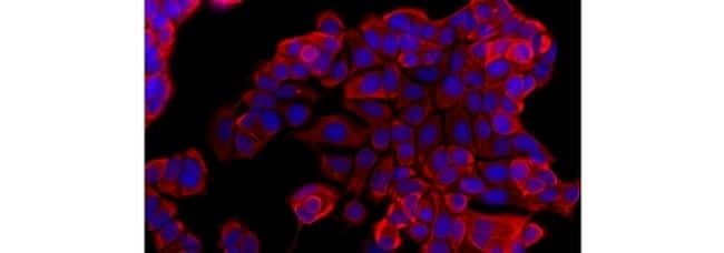
- Immunocytochemistry of fixed and permeabilized MCF7 cells using 1 µg/mL of Anti-Pan Cytokeratin (AE1/AE3) eFluor® 570. Nuclei are stained with DAPI.
Supportive validation
- Submitted by
- Invitrogen Antibodies (provider)
- Main image
- Experimental details
- NULL
- Submitted by
- Invitrogen Antibodies (provider)
- Main image
- Experimental details
- NULL
- Submitted by
- Invitrogen Antibodies (provider)
- Main image
- Experimental details
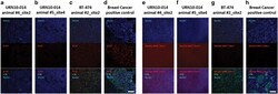
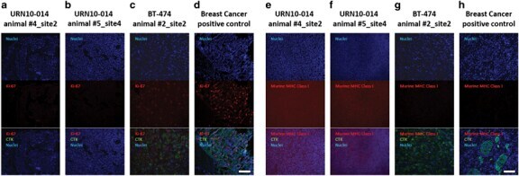
- Figure 2 Immunofluorescent staining for human-specific Ki-67, human-specific cytokeratin, and murine major histocompatibility complex (MHC) Class I. Nuclei were stained with DAPI. Observed neoplasms in the URN10-014 group were negative for human Ki-67+ and human-specific cytokeratin (columns a , b ), but stained positively for murine-specific MHC Class I ( e , f ). A BT474 xenograft ( c , g ) and a human metastatic breast cancer control ( d , h ) were positive for human cytokeratin and Ki-67, but negative for murine MHC Class I antigen. Scale bar (white)=100 mum.
- Submitted by
- Invitrogen Antibodies (provider)
- Main image
- Experimental details
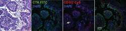
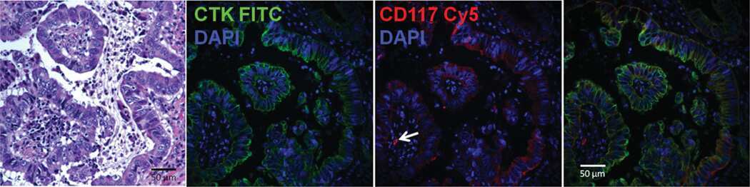
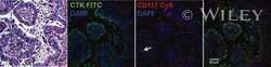
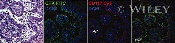
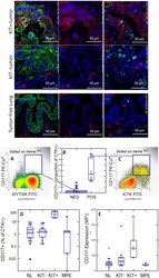
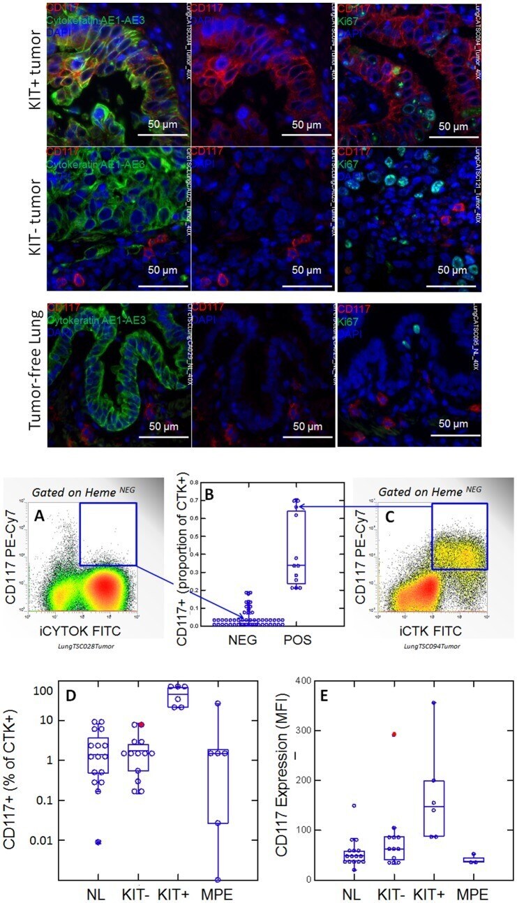
- Figure 1 CD117 expression in normal lung (NL) and NSCLC. Photomicrographs: Expression of CD117 and Ki-67 in NSC lung cancer and normal lung. The left columns show sections stained with CD117 (red), cytokeratin (green) and DAPI (blue). Sections in the center column show CD117 (red) and DAPI (blue) only, in order to reveal CD117 staining obscured by bright cytokeratin expression. Sections in the right column shows CD117 (red), the proliferation marker Ki67 (green) and DAPI (blue). Tumors were classified as KIT+ or negative on the basis of CD117 immunofluorescent staining of FFPE. In KIT+ tumors (top photomicrographs) CD117 (red stain, center and right panels) was expressed in virtually all cytokeratin+ tumor cells (green stain, left panels). Ki-67+ proliferating cells (green stain, right panels) were frequently seen among CD117+ tumor cells. In KIT negative tumors (center row of photomicrographs), only solitary CD117+ mast cells were detected (red stain, center and right panels). Proliferating Ki-67+ cells were frequent among cytokeratin+ CD117 negative tumor cells. Normal tumor-adjacent lung also appeared to lack CD117 expression among cytokeratin+ airway cells (bottom photomicrographs). Proliferating Ki-67+ cells were infrequent and confined to the basal layer of airway epithelium. When all NSCLC tumors are considered together, flow cytometry revealed bimodal CD117 expression (center panels A-C). Cells were gated on hematopoietic lineage negative singlet events with DNA conte
- Submitted by
- Invitrogen Antibodies (provider)
- Main image

- Experimental details
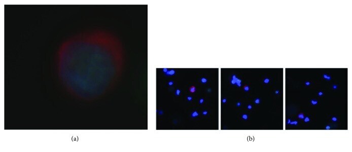
- Figure 1 (a) A circulating tumor cell prepared from a 7.5 ml blood sample from a 79-year-old male with no previous history of cancer. The cell is stained for cytokeratin (red) and for the cell nucleus (blue), typical of epithelial cells. Epithelial cells should not normally be present in the blood. The cell was negative for CD45, that is, not an immune cell. The cell nucleus has a large size typical of transcriptionally active cells, such as cancer cells, and the rounded shape of a cell in suspension, rather than the angular shape and cell sheath context of a normal solid tissue epithelium cell. (b) Three additional examples of circulating tumor cells stained for cytokeratin (red) and for the cell nucleus (blue). The lower magnification also shows the residual leucocytes surrounding the circulating tumor cells (blue nuclei, with no cytokeratin (red). The samples were enriched about 7500-fold for CTCs, with about 10,000 DAPI and CD45-positive leucocytes left in the sample after enrichment.
- Submitted by
- Invitrogen Antibodies (provider)
- Main image
- Experimental details
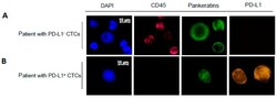
- Figure 2 Representative images of CTCs detected and subjected to immunostaining with DAPI, CD45, Pankeratins and PD-L1. Example images of CTCs from a patient with PD-L1 - CTCs ( A ) and PD-L1 + CTCs ( B ) are shown. The scale bar of 10 mum was applied to all pictures. ( C ) Number of CTCs and PD-L1 status isolated from blood samples from 38 patients (P1 through P38) with detectable PD-L1 status. ""P1"" stands for patient 1. Red bar represents the number of CK(+)/PD-L1(+) CTCs. Blue bar represents the number of CK(+)/PD-L1(-) CTCs.

- Submitted by
- Invitrogen Antibodies (provider)
- Main image
- Experimental details
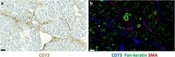
- Fig. 1 CD73 expression in normal pancreas. a Immunoperoxidase staining of normal pancreas for CD73 (brown). b Multicolour immunofluorescence staining of a consecutive section of normal pancreas for CD73 (blue), pan-cytokeratin (green) and alpha-smooth muscle actin (red). Bars, 50 mum
- Submitted by
- Invitrogen Antibodies (provider)
- Main image
- Experimental details
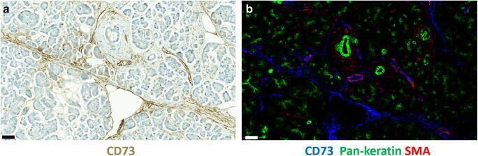
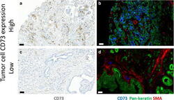
- Fig. 2 CD73 expression in adenocarcinoma of pancreas. Representative immunoperoxidase stainings (a and c) for CD73 (brown) and multicolour immunofluorescence stainings (b, d) for CD73 (blue), pan-cytokeratin (green) and alpha-smooth muscle actin (red). Bars, 50 mum

- Submitted by
- Invitrogen Antibodies (provider)
- Main image
- Experimental details
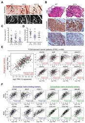
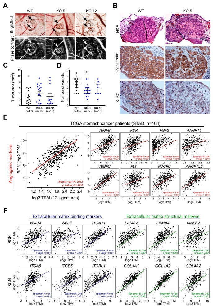
- Figure 6 Effect of biglycan in in vivo angiogenesis. ( A ) Representative images of tumors formed in the in vivo CAM by MKN74 cell models (WT and biglycan KO clones-KO.5 and KO.12). Phase contrast was used to better visualize tumor foci and vessels. MKN74 form multiple tumor foci (arrows/circles). Magnification at 20x. ( B ) Histological images of the formed tumors, cytokeratin staining confirming the presence of human epithelial tumors in the CAM. Ki-67 expression analysis was used to assess tumor aggressiveness. Histologically, tumors formed by KO cells present a less cohesive-like tumor mass with increased extracellular matrix stiffness. Hematoxilin & Eosin (H&E) staining images at 100X magnification, cytokeration and Ki-67 images at 200x magnification. ( C ) Quantification of the tumor area (mm 2 ) with no significant differences in the tumors being derived from the different cell lines. ( D ) Number of new vessels (less than 20 mum in diameter) formed towards the inoculation site. biglycan KO inoculated cells showed less capacity to form new vessels when compared to WT biglycan-positive tumors. ( E ) In silico gene analysis in GC tissues samples (TCGA, n = 408) showing that BGN was strongly positively correlated with angiogenic markers ( VEGFB, VEGFC, KDR, FLT1, FGF2, PDGFC, ANGPT1, ANGPT2, ANGPTL2, ANGPTL1, ANGPTL4, and ANGPTL7 ) . ( F ) In silico analysis demonstrating that BGN is positively correlated with ECM binding ( VCAM1, SELE, ITGA11, ITGA5, ITGB5, ITGBL1 ) and
- Submitted by
- Invitrogen Antibodies (provider)
- Main image
- Experimental details
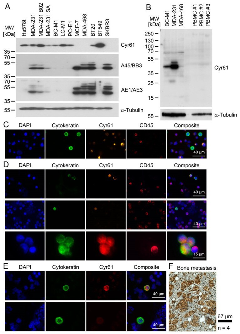
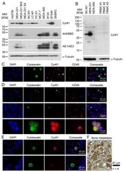
- Figure 5 Cyr61 detection in breast cancer cells. ( A ) Comparison of the cytoplasmic Cyr61 levels with cytokeratin levels, as analysed by the pan-cytokeratin antibody cocktails A45/BB3 and AE1/AE3 by Western blot analysis. ( B ) A comparison of the Cyr61 levels in the peripheral blood mononuclear cells (PBMC) of healthy women with the Cyr61 levels in breast cancer cell lines. ( C ) Cyr61 detection in BC-M1 and MDA-MB-468 spiked into blood samples from healthy women by immunocytochemical double staining. ( D ) Cyr61 detection in CTC from the peripheral blood of breast cancer patients (details: Table 1 ). ( E ) Detection of Cyr61 in the DTC from the bone marrow of breast cancer patients. The upper row shows a Cyr61-positive DTC, and the bottom row shows a Cyr61-negative DTC. ( F ) An immunohistochemical Cyr61 detection in the bone metastases of breast cancer patients. ( C - E ) The composite images are overlays of the Cytokeratin, Cyr61, Dapi and CD45 (if applied) signals, n biol : 3 ( A , C , E ), 4 ( F ).