Explore
 Validate
 Learn
Learn Western blot
Antibody data
- Antibody Data
- Antigen structure
- References [0]
- Comments [0]
- Validations
- Western blot [3]
- Immunocytochemistry [3]
- Chromatin Immunoprecipitation [1]
Submit
Validation data
Reference
Comment
Report error
- Product number
- PA5-40091 - Provider product page

- Provider
- Invitrogen Antibodies
- Product name
- HP1 alpha/beta/gamma Polyclonal Antibody
- Antibody type
- Polyclonal
- Antigen
- Other
- Reactivity
- Human, Mouse
- Host
- Rabbit
- Isotype
- IgG
- Vial size
- 50 µg
- Concentration
- 2 mg/mL
- Storage
- -20°C or -80°C if preferred
No comments: Submit comment
Supportive validation
- Submitted by
- Invitrogen Antibodies (provider)
- Main image

- Experimental details
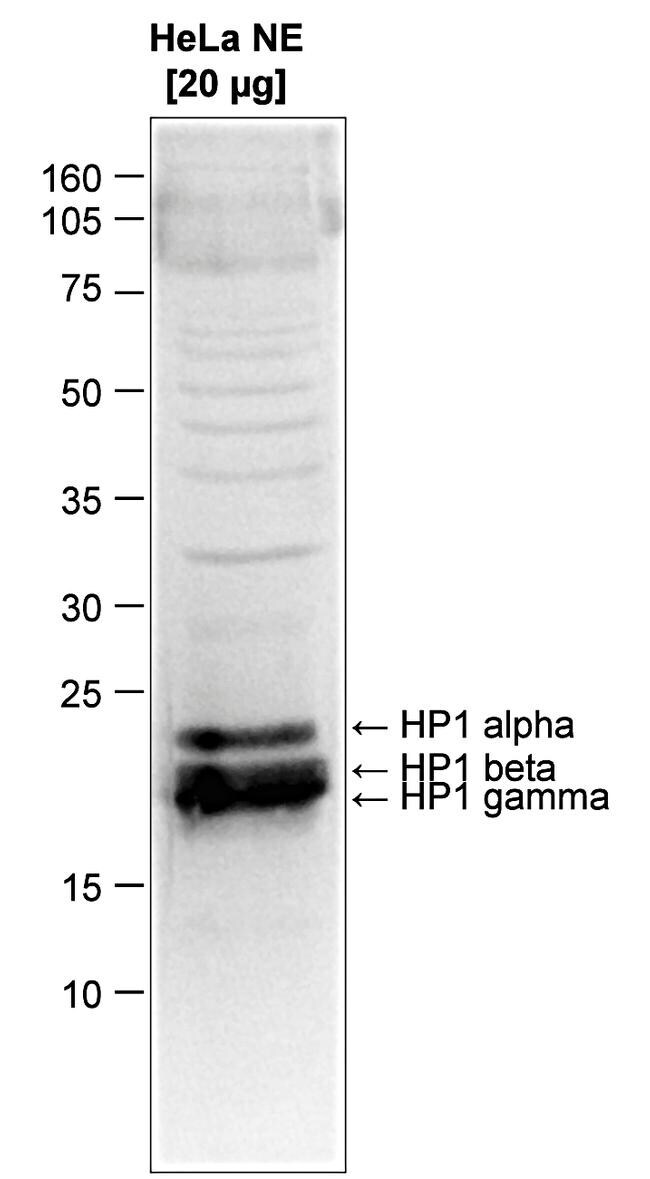
- Western Blot was performed on nuclear extracts from HeLa cells (20 µg) using a HP1 alpha + beta + gamma polyclonal antibody (Product # PA5-40091) at a dilution of 1:1,000 in TBS-Tween containing 5% skimmed milk (Figure 1). The molecular weight marker (kDa) is shown on the left; the expected location of HP1-alpha, HP1-beta and HP1-gamma is indicated on the right.
- Submitted by
- Invitrogen Antibodies (provider)
- Main image
- Experimental details
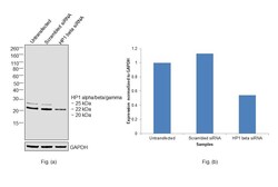
- Knockdown of Chromobox protein homolog 5 and 1 was achieved by transfecting HeLa with Chromobox protein homolog 1 specific siRNAs (Silencer® select Product # S21549, S21550). Western blot analysis (Fig. a) was performed using Nuclear enriched extracts from the Chromobox protein homolog 5 and 1 knockdown cells (lane 3), non-targeting scrambled siRNA transfected cells (lane 2) and untransfected cells (lane 1). The blot was probed with HP1 alpha/beta/gamma Polyclonal Antibody (Product # PA5-40091, 1:1000 dilution) and Goat anti-Rabbit IgG (H+L) Superclonal™ Recombinant Secondary Antibody, HRP (Product # A27036, 1:20000 dilution). Densitometric analysis of this western blot is shown in histogram (Fig. b). Decrease in signal upon siRNA mediated knock down confirms that antibody is specific to Chromobox protein homolog 5 and 1.

- Submitted by
- Invitrogen Antibodies (provider)
- Main image
- Experimental details
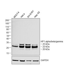
- Western blot was performed using Anti-HP1 alpha/beta/gamma Polyclonal Antibody (Product # PA5-40091) and a 20, 22, 25 kDa bands corresponding to Chromobox protein homolog 3, 1 and 5 respectively were observed across tested cell lines. Nuclear enriched extracts (40 µg lysate) of MOLT-4 (Lane 1), HeLa (Lane 2), SH-SY5Y (Lane 3), Hep G2 (Lane 4) were electrophoresed using NuPAGE™ 12% Bis-Tris Protein Gel (Product # NP0341BOX). Resolved proteins were then transferred onto a nitrocellulose membrane (Product # IB23001) by iBlot® 2 Dry Blotting System (Product # IB21001). The blot was probed with the primary antibody (1:1000 dilution) and detected by chemiluminescence with Goat anti-Rabbit IgG (H+L) Superclonal™ Recombinant Secondary Antibody, HRP (Product # A27036,1:20000 dilution using the iBright™ FL1500 Imaging System (Product # A44115). Chemiluminescent detection was performed using Novex® ECL Chemiluminescent Substrate Reagent Kit (Product # WP20005).

Supportive validation
- Submitted by
- Invitrogen Antibodies (provider)
- Main image

- Experimental details
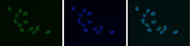
- Immunofluorescent detection of HP1 alpha + beta + gamma in HeLa cells using a HP1 alpha + beta + gamma polyclonal antibody (Product # PA5-40091). Cells were fixed with 4% formaldehyde for 10' and blocked with PBS/Triton X-100 containing 5% normal goat serum and 1% BSA. The cells were immunofluorescently labelled with the HP1-alpha, -beta, -gamma antibody (left) at a dilution of 1:500 in blocking solution followed by detection using an anti-rabbit antibody conjugated to AlexaFluor 488. The middle panel shows staining of the nuclei with DAPI. A merge of the two stainings is shown on the right.
- Submitted by
- Invitrogen Antibodies (provider)
- Main image

- Experimental details
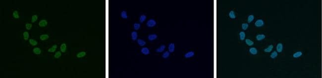
- Immunofluorescent detection of HP1 alpha + beta + gamma in HeLa cells using a HP1 alpha + beta + gamma polyclonal antibody (Product # PA5-40091). Cells were fixed with 4% formaldehyde for 10' and blocked with PBS/Triton X-100 containing 5% normal goat serum and 1% BSA. The cells were immunofluorescently labelled with the HP1-alpha, -beta, -gamma antibody (left) at a dilution of 1:500 in blocking solution followed by detection using an anti-rabbit antibody conjugated to AlexaFluor 488. The middle panel shows staining of the nuclei with DAPI. A merge of the two stainings is shown on the right.
- Submitted by
- Invitrogen Antibodies (provider)
- Main image

- Experimental details
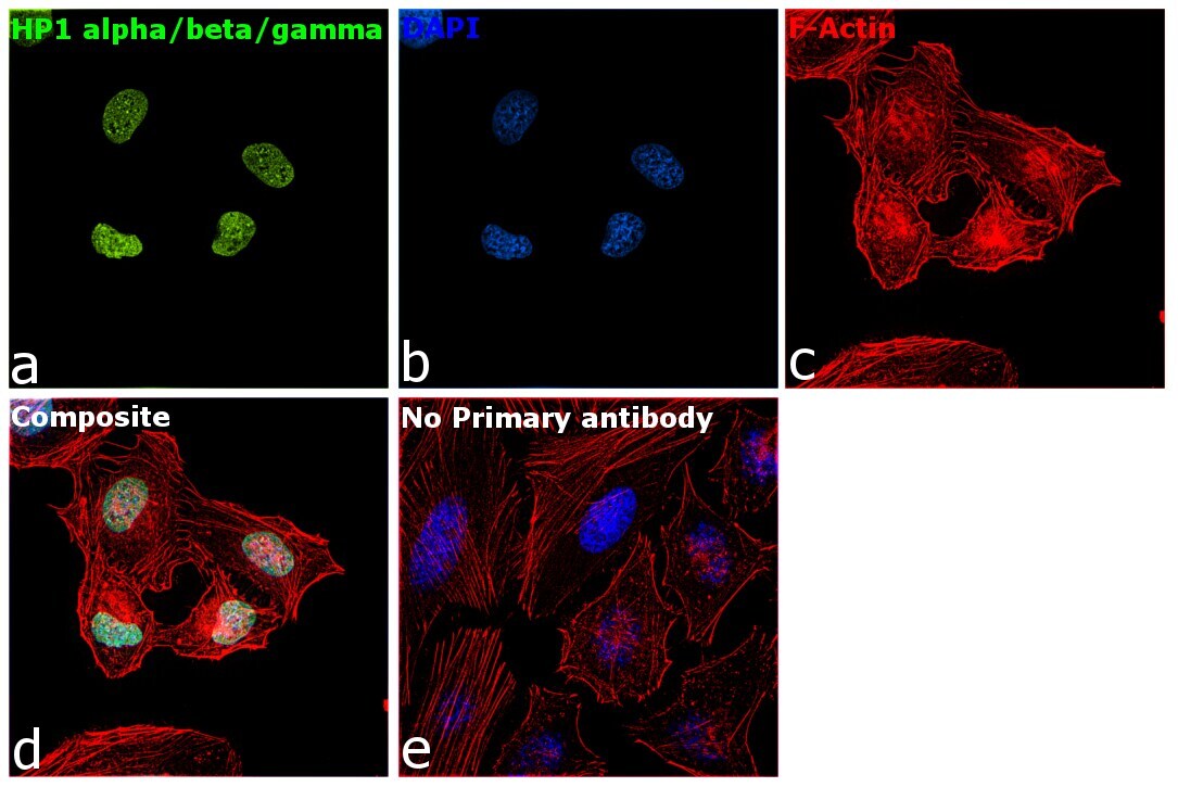
- Immunofluorescence analysis of Chromobox protein homolog 5 was performed using 70% confluent log phase HeLa cells. The cells were fixed with 4% paraformaldehyde for 5 minutes, permeabilized with 0.1% Triton™ X-100 for 10 minutes, and blocked with 2% BSA for 45 minutes at room temperature. The cells were labeled with HP1 alpha/beta/gamma Polyclonal Antibody (Product # PA5-40091) at 1:100 dilution in 0.1% BSA, incubated at 4 degree celsius overnight and then labeled with Donkey anti-Rabbit IgG (H+L) Highly Cross-Adsorbed Secondary Antibody, Alexa Fluor Plus 488 (Product # A32790), (1:2000 dilution), for 45 minutes at room temperature (Panel a: Green). Nuclei (Panel b:Blue) were stained with ProLong™ Diamond Antifade Mountant with DAPI (Product # P36962). F-actin (Panel c: Red) was stained with Rhodamine Phalloidin (Product # R415, 1:300). Panel d represents the merged image showing nuclear localization. Panel e represents control cells with no primary antibody to assess background. The images were captured at 60X magnification.
Supportive validation
- Submitted by
- Invitrogen Antibodies (provider)
- Main image
- Experimental details
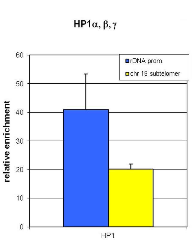
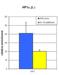
- ChIP assays were performed on NIH3T3 cells using 4 µg of HP1 alpha + beta + gamma polyclonal antibody (Product # PA5-40091). QPCR was performed on the IP'd DNA with optimized primer sets for the rDNA promoter and for a subtelomeric sequence of chromosome 19. Figure 1 shows the relative enrichment as compared to a no antibody negative control ChIP.