Explore
 Validate
 Learn
Learn Immunohistochemistry
 Flow cytometry
Antibody data
- Antibody Data
- Antigen structure
- References [2]
- Comments [0]
- Validations
- Flow cytometry [1]
Submit
Validation data
Reference
Comment
Report error
- Product number
- 14-0519-80 - Provider product page

- Provider
- Invitrogen Antibodies
- Product name
- Anti-CD51/CD61 (Integrin alpha v beta 3) Monoclonal Antibody (23C6), eBioscience™
- Antibody type
- Monoclonal
- Antigen
- Other
- Description
- Description: The 23C6 monoclonal antibody reacts with the human CD51/CD61 dimer, also known as the integrin alphav/beta3. CD51, an ~120 kDa surface molecule can also non-covalently associate with other beta subunits of the integrin family including beta1 (CD29), beta5 and beta6 to form receptors for extracellular matrix components. Heterodimers of CD51/CD61 are expressed by melanoma cells, endothelial cells and osteoclasts and at very low levels by platelets. The CD51/CD61 complex mediates adhesion to fibrinogen, fibronectin, vitronectin and thrombospondin. Applications Reported: The 23C6 antibody has been reported for use in flow cytometric analysis, and immunohistochemical staining of frozen tissue sections. 23C6 has also been reported in blocking of some adhesive processes. (Please use Functional Grade purified 23C6 in functional assays.). Applications Tested: The 23C6 antibody has been tested by flow cytometric analysis of human melanoma cell line and peripheral blood. This can be used at less than or equal to 1 µg per test. A test is defined as the amount (µg) of antibody that will stain a cell sample in a final volume of 100 µL. Cell number should be determined empirically but can range from 10^5 to 10^8 cells/test. It is recommended that the antibody be carefully titrated for optimal performance in the assay of interest. Purity: Greater than 90%, as determined by SDS-PAGE. Aggregation: Less than 10%, as determined by HPLC. Filtration: 0.2 µm post-manufacturing filtered.
- Reactivity
- Human
- Host
- Mouse
- Isotype
- IgG
- Antibody clone number
- 23C6
- Vial size
- 25 µg
- Concentration
- 0.5 mg/mL
- Storage
- 4° C
Submitted references A hyperspectral method to assay the microphysiological fates of nanomaterials in histological samples.
The matricellular protein CCN1 mediates neutrophil efferocytosis in cutaneous wound healing.
SoRelle ED, Liba O, Campbell JL, Dalal R, Zavaleta CL, de la Zerda A
eLife 2016 Aug 18;5
eLife 2016 Aug 18;5
The matricellular protein CCN1 mediates neutrophil efferocytosis in cutaneous wound healing.
Jun JI, Kim KH, Lau LF
Nature communications 2015 Jun 16;6:7386
Nature communications 2015 Jun 16;6:7386
No comments: Submit comment
Supportive validation
- Submitted by
- Invitrogen Antibodies (provider)
- Main image

- Experimental details
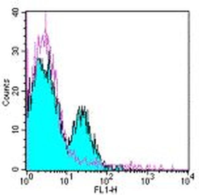
- Staining of platelets with 0.5 µg of Mouse IgG1 Isotype Control Purified (Product # 14-4714-82) (open histogram) or 0.5 µg of Anti-Human CD51/CD61 (Integrin alpha/beta 3) Purified followed by Anti-Mouse IgG FITC (Product # 11-4011-85) (filled histogram).