 Explore
 Validate
 Learn
Learn Western blot
 Immunocytochemistry
Antibody data
- Antibody Data
- Antigen structure
- References [0]
- Comments [0]
- Validations
- Immunocytochemistry [1]
- Other assay [1]
Submit
Validation data
Reference
Comment
Report error
- Product number
- PA1-1000 - Provider product page

- Provider
- Invitrogen Antibodies
- Product name
- GNAI1/GNAI2 Polyclonal Antibody
- Antibody type
- Polyclonal
- Antigen
- Synthetic peptide
- Description
- PA1-1000 detects G protein alpha inhibitor 1/2 from human, rat, and mouse tissues and cells. PA1-1000 has been successfully used in Immunoprecipitation and Western blot procedures. By Western blot, this antibody detects an ~38 kDa protein representing G protein alpha inhibitor 1/2 from HeLa cell lysate. The PA1-1000 immunogen is a synthetic peptide corresponding to residues K(346) N N L K D C G L F(355) of human G Protein alpha Inhibitor 1/2. This sequence is completely conserved in human, rat, guinea pig, hamster, bovine, hydra, octopus, Xenopus, chicken, canine, spiny dogfish, great pond snail, starfish, and American lobster G protein alpha inhibitor 1/2. PA1-1000 immunizing peptide (Cat. # PEP-129) is available for use in neutralization and control procedures.
- Reactivity
- Human, Mouse, Rat
- Host
- Rabbit
- Isotype
- IgG
- Vial size
- 100 µg
- Concentration
- 1 mg/mL
- Storage
- -20° C, Avoid Freeze/Thaw Cycles
No comments: Submit comment
Supportive validation
- Submitted by
- Invitrogen Antibodies (provider)
- Main image
- Experimental details
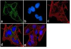
- Immunofluorescence analysis of G Protein alpha Inhibitor 1 & 2 was performed using 70% confluent log phase SH-SY5Y cells. The cells were fixed with 4% paraformaldehyde for 10 minutes, permeabilized with 0.1% Triton™ X-100 for 10 minutes, and blocked with 1% BSA for 1 hour at room temperature. The cells were labeled with GNAI1/GNAI2 Rabbit Polyclonal Antibody (Product # PA1-1000) at 2 µg/mL in 0.1% BSA and incubated for 3 hours at room temperature and then labeled with Goat anti-Rabbit IgG (H+L) Superclonal™ Secondary Antibody, Alexa Fluor® 488 conjugate (Product # A27034) at a dilution of 1:2000 for 45 minutes at room temperature (Panel a: green). Nuclei (Panel b: blue) were stained with SlowFade® Gold Antifade Mountant with DAPI (Product # S36938). F-actin (Panel c: red) was stained with Alexa Fluor® 555 Rhodamine Phalloidin (Product # R415, 1:300). Panel d represents the merged image showing cytoplasmic and membranous localization. Panel e shows the no primary antibody control. The images were captured at 60X magnification.

Supportive validation
- Submitted by
- Invitrogen Antibodies (provider)
- Main image
- Experimental details
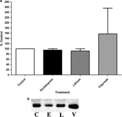
- Figure 2 Gialpha membrane compartmentalization is not altered by chronic antidepressant or lithium treatment. Purified lipid rafts isolated using sucrose gradient flotation revealed that neither chronic antidepressant treatment nor chronic lithium treatment move Gialpha out of lipid rafts. a The figure shows the percentage of change in Gialpha protein in the lipid raft membrane fractions compared to control (n = 4 for control, escitalopram, and lithium; n = 3 for valproate). Data were analyzed by one-way ANOVA followed by Tukey's multiple comparison test for post hoc comparison of means. Data are represented as mean +- SEM b A representative immunoblot of lipid raft Gsalpha from the various treatment paradigms as explained in "" Methods "". C control, E escitalopram, L lithium, V valproic acid.
