Explore
 Validate
 Learn
Learn Western blot
 Immunocytochemistry
Antibody data
- Antibody Data
- Antigen structure
- References [2]
- Comments [0]
- Validations
- Immunocytochemistry [1]
Submit
Validation data
Reference
Comment
Report error
- Product number
- HPA027141 - Provider product page

- Provider
- Atlas Antibodies
- Proper citation
- Atlas Antibodies Cat#HPA027141, RRID:AB_1844619
- Product name
- Anti-ADPRHL2
- Antibody type
- Polyclonal
- Description
- Polyclonal Antibody against Human ADPRHL2, Gene description: ADP-ribosylhydrolase like 2, Alternative Gene Names: ARH3, FLJ20446, Validated applications: ICC, IHC, WB, Uniprot ID: Q9NX46, Storage: Store at +4°C for short term storage. Long time storage is recommended at -20°C.
- Reactivity
- Human
- Host
- Rabbit
- Conjugate
- Unconjugated
- Isotype
- IgG
- Vial size
- 100 µl
- Concentration
- 0.1 mg/ml
- Storage
- Store at +4°C for short term storage. Long time storage is recommended at -20°C.
- Handling
- The antibody solution should be gently mixed before use.
Submitted references Bi-allelic ADPRHL2 Mutations Cause Neurodegeneration with Developmental Delay, Ataxia, and Axonal Neuropathy
ADP-ribosylhydrolase 3 (ARH3), Not Poly(ADP-ribose) Glycohydrolase (PARG) Isoforms, Is Responsible for Degradation of Mitochondrial Matrix-associated Poly(ADP-ribose)
Danhauser K, Alhaddad B, Makowski C, Piekutowska-Abramczuk D, Syrbe S, Gomez-Ospina N, Manning M, Kostera-Pruszczyk A, Krahn-Peper C, Berutti R, Kovács-Nagy R, Gusic M, Graf E, Laugwitz L, Röblitz M, Wroblewski A, Hartmann H, Das A, Bültmann E, Fang F, Xu M, Schatz U, Karall D, Zellner H, Haberlandt E, Feichtinger R, Mayr J, Meitinger T, Prokisch H, Strom T, Płoski R, Hoffmann G, Pronicki M, Bonnen P, Morlot S, Haack T
The American Journal of Human Genetics 2018;103(5):817-825
The American Journal of Human Genetics 2018;103(5):817-825
ADP-ribosylhydrolase 3 (ARH3), Not Poly(ADP-ribose) Glycohydrolase (PARG) Isoforms, Is Responsible for Degradation of Mitochondrial Matrix-associated Poly(ADP-ribose)
Niere M, Mashimo M, Agledal L, Dölle C, Kasamatsu A, Kato J, Moss J, Ziegler M
Journal of Biological Chemistry 2012;287(20):16088-16102
Journal of Biological Chemistry 2012;287(20):16088-16102
No comments: Submit comment
Supportive validation
- Submitted by
- Atlas Antibodies (provider)
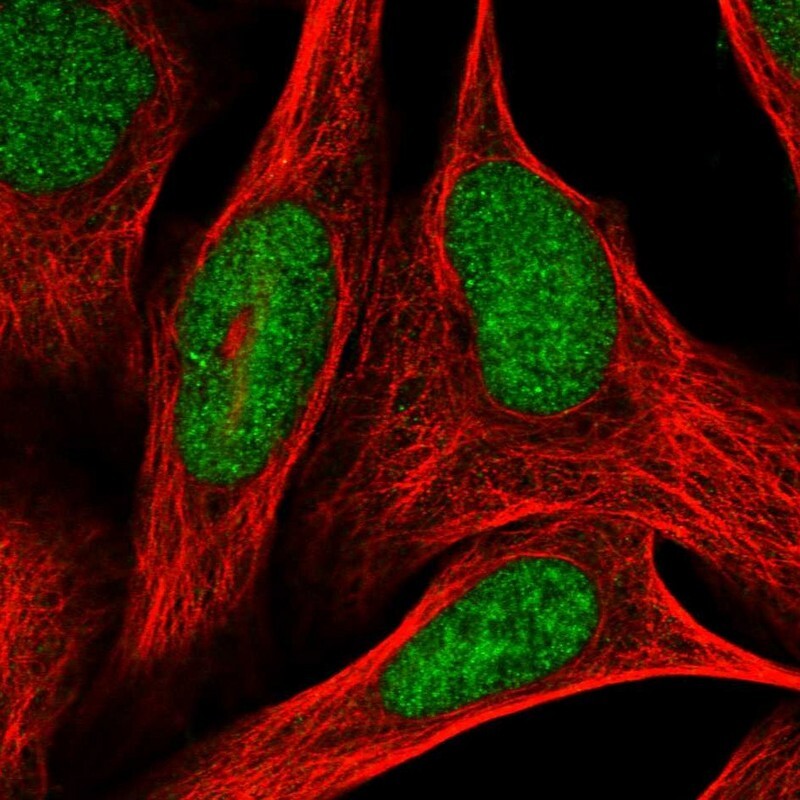
- Main image

- Experimental details
- Immunofluorescent staining of human cell line U-2 OS shows localization to nucleoplasm.
- Sample type
- Human