Explore
 Validate
 Learn
Learn Western blot
Antibody data
- Antibody Data
- Antigen structure
- References [2]
- Comments [0]
- Validations
- Western blot [4]
- Immunohistochemistry [6]
Submit
Validation data
Reference
Comment
Report error
- Product number
- NBP1-85937 - Provider product page

- Provider
- Novus Biologicals
- Proper citation
- Novus Cat#NBP1-85937, RRID:AB_11012461
- Product name
- Rabbit Polyclonal DARS Antibody
- Antibody type
- Polyclonal
- Description
- Immunogen affinity purified. Specificity of human DARS antibody verified on a Protein Array containing target protein plus 383 other non-specific proteins.
- Reactivity
- Human, Mouse
- Host
- Rabbit
- Isotype
- IgG
- Vial size
- 0.1 ml
- Storage
- Store at 4C short term. Aliquot and store at -20C long term. Avoid freeze-thaw cycles.
Submitted references Expression Pattern of the Aspartyl-tRNA Synthetase DARS in the Human Brain.
In vivocharacterization of the aspartyl-tRNA synthetase DARS: Homing in on the leukodystrophy HBSL.
Fröhlich D, Suchowerska AK, Voss C, He R, Wolvetang E, von Jonquieres G, Simons C, Fath T, Housley GD, Klugmann M
Frontiers in molecular neuroscience 2018;11:81
Frontiers in molecular neuroscience 2018;11:81
In vivocharacterization of the aspartyl-tRNA synthetase DARS: Homing in on the leukodystrophy HBSL.
Fröhlich D, Suchowerska AK, Spencer ZH, von Jonquieres G, Klugmann CB, Bongers A, Delerue F, Stefen H, Ittner LM, Fath T, Housley GD, Klugmann M
Neurobiology of disease 2017 Jan;97(Pt A):24-35
Neurobiology of disease 2017 Jan;97(Pt A):24-35
No comments: Submit comment
Supportive validation
- Submitted by
- Novus Biologicals (provider)
- Main image
- Experimental details
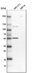
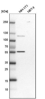
- Western Blot: DARS Antibody [NBP1-85937] - Analysis in mouse cell line NIH-3T3 and rat cell line NBT-II.
- Submitted by
- Novus Biologicals (provider)
- Main image
- Experimental details
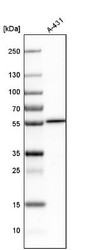
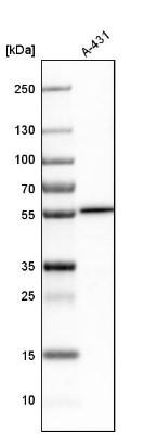
- Western Blot: DARS Antibody [NBP1-85937] - Analysis in human cell line A-431.
- Submitted by
- Novus Biologicals (provider)
- Main image
- Experimental details
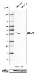
- Western Blot: DARS Antibody [NBP1-85937] - Analysis in A-431 cells transfected with control siRNA, target specific siRNA probe #1, using Anti-DARS antibody. Remaining relative intensity is presented. Loading control: Anti-GAPDH.

- Submitted by
- Novus Biologicals (provider)
- Main image
- Experimental details
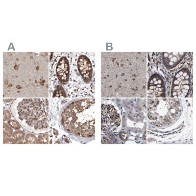
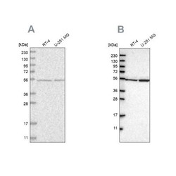
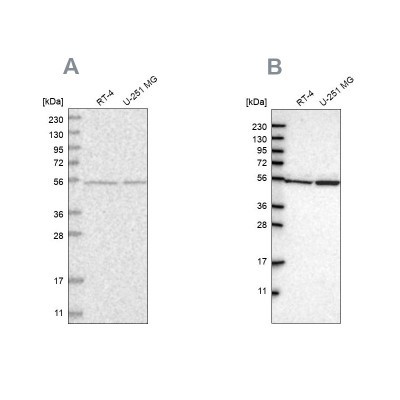
- Western Blot: DARS Antibody [NBP1-85937] - Analysis using Anti-DARS antibody NBP1-85937 (A) shows similar pattern to independent antibody NBP1-86027 (B).
Supportive validation
- Submitted by
- Novus Biologicals (provider)
- Main image

- Experimental details
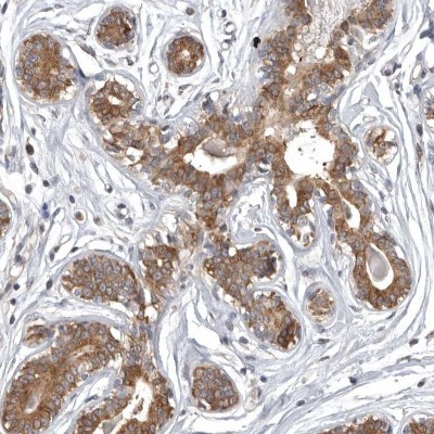
- Immunohistochemistry-Paraffin: DARS Antibody [NBP1-85937] - Staining of human breast shows cytoplasmic positivity in glandular cells.
- Submitted by
- Novus Biologicals (provider)
- Main image
- Experimental details
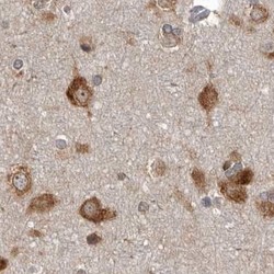
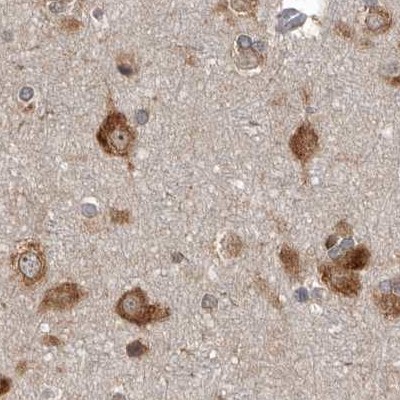
- Immunohistochemistry-Paraffin: DARS Antibody [NBP1-85937] - Staining of human cerebral cortex.
- Submitted by
- Novus Biologicals (provider)
- Main image
- Experimental details
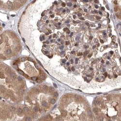
- Immunohistochemistry-Paraffin: DARS Antibody [NBP1-85937] - Staining of human kidney.

- Submitted by
- Novus Biologicals (provider)
- Main image
- Experimental details
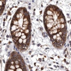
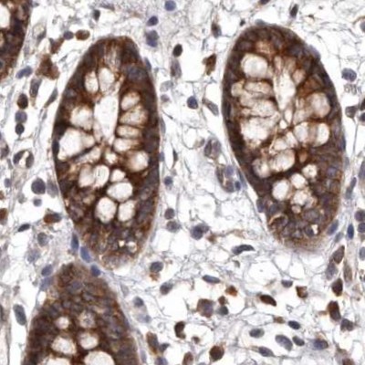
- Immunohistochemistry-Paraffin: DARS Antibody [NBP1-85937] - Staining of human colon.
- Submitted by
- Novus Biologicals (provider)
- Main image
- Experimental details
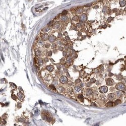
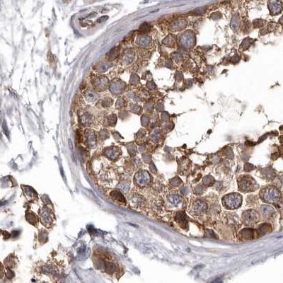
- Immunohistochemistry-Paraffin: DARS Antibody [NBP1-85937] - Staining of human testis.
- Submitted by
- Novus Biologicals (provider)
- Main image

- Experimental details
- Immunohistochemistry-Paraffin: DARS Antibody [NBP1-85937] - Staining of human cerebral cortex, colon, kidney and testis using Anti-DARS antibody NBP1-85937 (A) shows similar protein distribution across tissues to independent antibody NBP1-86027 (B).