Explore
 Validate
 Learn
Learn Western blot
Antibody data
- Antibody Data
- Antigen structure
- References [0]
- Comments [0]
- Validations
- Western blot [1]
- ELISA [1]
- Immunohistochemistry [2]
- Other assay [4]
Submit
Validation data
Reference
Comment
Report error
- Product number
- MA1-141 - Provider product page

- Provider
- Invitrogen Antibodies
- Product name
- Collagen I Monoclonal Antibody (5D8-G9)
- Antibody type
- Monoclonal
- Antigen
- Purifed from natural sources
- Description
- MA1-141 detects collagen I from bovine, human, and porcine. This antibody will not cross react with collagen 2-11, or detect thermally denatured collagen. Western blot analysis of recombinant bovine collagen I using MA1-141 detected a predominant band around 270kD likely corresponding to the alpha-1 dimer and a fainter band at a higher MW likely corresponding to the alpha-1/alpha-2 trimer. MA1-141 has been successfully used in ELISA, immunofluorescence, immunohistochemistry (frozen and paraffin), and Western blot procedures. The epitope is sensitive to routine formalin fixation and paraffin embedding. Strong staining of connective tissue fibres is seen in acetone-fixed or unfixed frozen sections.
- Reactivity
- Human, Bovine, Porcine
- Host
- Mouse
- Isotype
- IgG
- Antibody clone number
- 5D8-G9
- Vial size
- 100 µg
- Concentration
- 1 mg/mL
- Storage
- -20°C
No comments: Submit comment
Supportive validation
- Submitted by
- Invitrogen Antibodies (provider)
- Main image
- Experimental details
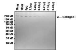
- Western blot analysis of collagen I was performed by loading the indicated amounts of bovine collagen I onto a 4-12% Bis-Tris polyacrylamide gel. Proteins were transferred to a nitrocellulose membrane and blocked with 5% BSA in TBST for at least 1 hour. The membrane was probed with a collagen I monoclonal antibody (Product # MA1-141) at a dilution of 1:1000 overnight at 4°C on a rocking platform, washed in TBST, and probed with an HRP-conjugated goat anti-mouse IgG secondary antibody (Product # 31430) at a dilution of 1:20,000 for 1 hour. Chemiluminescent detection was performed using SuperSignal West Dura (Product # 34076).
Supportive validation
- Submitted by
- Invitrogen Antibodies (provider)
- Main image
- Experimental details
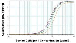
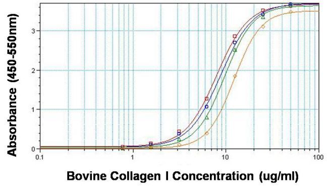
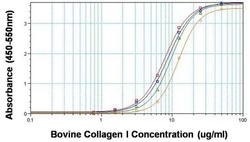
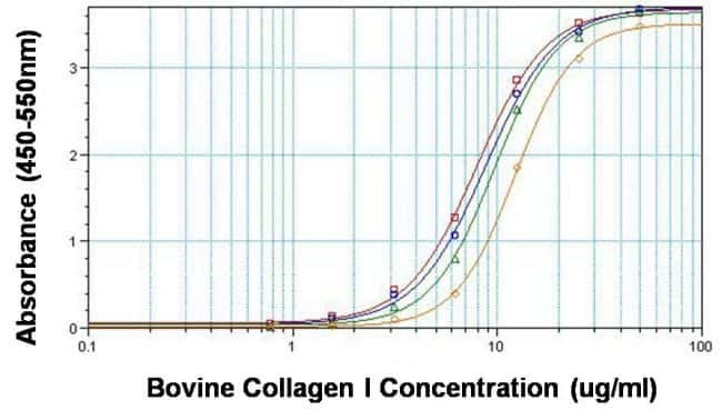
- Direct ELISA analysis of collagen I was performed by coating 100 µL per well of bovine collagen I in twelve replicates at 50, 25, 12.5, 6.25, 3.12, 1.56, 0.781 and 0 µg/mL across a clear, 96-well plate and incubating overnight at room temperature. The coating solution was decanted and the plate was blocked with 5% BSA in TBST for at least 2 hours. The plate was washed with TBST and then incubated with 100 µL per well of collagen I monoclonal antibody (Product # MA1-141) at 50 µg/mL (red line), 10 µg/mL (blue line), 2 µg/mL (green line) and 0.4 µg/mL (orange line) in blocking buffer for 1 hour at room temperature. The plate was washed and incubated with 100 µL per well of HRP-conjugated goat anti-mouse IgG secondary antibody (Product # 31430) in all test wells at 1:10,000 for 30 minutes at room temperature. The plate was again washed in TBST and detection was performed using ultra TMB substrate (Product # 34028) for 30 minutes at room temperature in the dark. The reaction was then stopped with 0.16M sulfuric acid and absorbance was read at 450-550 nm.
Supportive validation
- Submitted by
- Invitrogen Antibodies (provider)
- Main image
- Experimental details
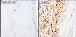
- Immunohistochemistry analysis of Collagen I was performed on porcine skin tissue. To expose target proteins, antigen retrieval was performed by microwaving tissues for 8-15 minutes in 10mM sodium citrate buffer (pH 6.0). Following antigen retrieval, tissues were blocked in 3% hydrogen peroxide-methanol for 15 min at room temperature, washed with deionized water and PBS, and then probed with a Collagen I monoclonal antibody (Product # MA1-141) diluted 1:20 in 3% BSA-PBS (right panel) or incubated with buffer alone not containing primary antibody as a negative control (left panel), overnight at 4°C in a humidified chamber. Tissues were washed extensively in PBST and detection was performed using an HRP-conjugated secondary antibody followed by colorimetric detection using a DAB kit. Tissues were counterstained with hematoxylin and dehydrated with ethanol and xylene to prep for mounting.
- Submitted by
- Invitrogen Antibodies (provider)
- Main image
- Experimental details
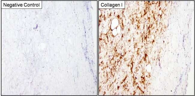
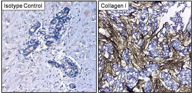
- Immunohistochemistry analysis of Collagen I was performed on frozen human breast carcinoma tissue. Tissues were probed with a Collagen I monoclonal antibody (Product # MA1-141, right panel) or mouse IgG isotype control (left panel) at a dilution of 1:500 for 1 hour at room temperature. Tissues were washed, and detection was performed using an HRP-conjugated universal detection reagent followed by DAB substrate. Tissues were counterstained and prepped for mounting before visualization by light microscopy.
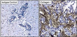
Supportive validation
- Submitted by
- Invitrogen Antibodies (provider)
- Main image
- Experimental details
- NULL

- Submitted by
- Invitrogen Antibodies (provider)
- Main image
- Experimental details
- NULL

- Submitted by
- Invitrogen Antibodies (provider)
- Main image
- Experimental details
- Direct ELISA analysis of collagen I was performed by coating 100 µL per well of bovine collagen I in twelve replicates at 50, 25, 12.5, 6.25, 3.12, 1.56, 0.781 and 0 µg/mL across a clear, 96-well plate and incubating overnight at room temperature. The coating solution was decanted and the plate was blocked with 5% BSA in TBST for at least 2 hours. The plate was washed with TBST and then incubated with 100 µL per well of collagen I monoclonal antibody (Product # MA1-141) at 50 µg/mL (red line), 10 µg/mL (blue line), 2 µg/mL (green line) and 0.4 µg/mL (orange line) in blocking buffer for 1 hour at room temperature. The plate was washed and incubated with 100 µL per well of HRP-conjugated goat anti-mouse IgG secondary antibody (Product # 31430) in all test wells at 1:10,000 for 30 minutes at room temperature. The plate was again washed in TBST and detection was performed using ultra TMB substrate (Product # 34028) for 30 minutes at room temperature in the dark. The reaction was then stopped with 0.16M sulfuric acid and absorbance was read at 450-550 nm.
- Submitted by
- Invitrogen Antibodies (provider)
- Main image

- Experimental details
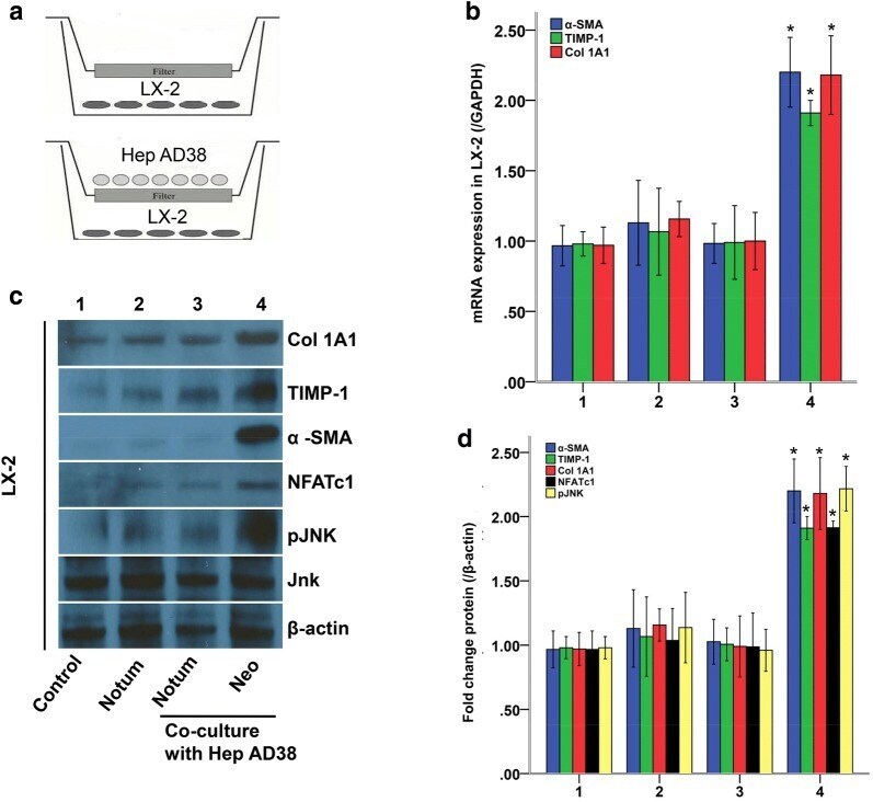
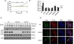
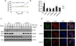
- Fig. 3 Notum inhibited liver fibrosis gene expression in LX-2 cells with HBV exposure. a Cell co-culture model. b LX-2 cells were co-cultured with Hep AD38 cells and transferred with Notum. alpha-SMA, TIMP-1 and Col 1A1 mRNA expressions in LX-2 cells were measured by qPCR. c , d alpha-SMA, TIMP-1 and Col 1A1 protein expressions in LX-2 cells were measured by Western-blot. * P < 0.05 compared with mock. All experiments were tried 3 times