 Explore
 Validate
 Learn
Learn Western blot
 Immunocytochemistry
Antibody data
- Antibody Data
- Antigen structure
- References [1]
- Comments [0]
- Validations
- Western blot [3]
- Immunohistochemistry [3]
Submit
Validation data
Reference
Comment
Report error
- Product number
- NBP1-33610 - Provider product page

- Provider
- Novus Biologicals
- Proper citation
- Novus Cat#NBP1-33610, RRID:AB_2134915
- Product name
- Rabbit Polyclonal Loricrin Antibody
- Antibody type
- Polyclonal
- Description
- Immunogen affinity purified.
- Reactivity
- Human, Mouse
- Host
- Rabbit
- Isotype
- IgG
- Vial size
- 0.1 ml
- Storage
- Aliquot and store at -20C or -80C. Avoid freeze-thaw cycles.
Submitted references Immunotopographical Differences of Human Skin.
Béke G, Dajnoki Z, Kapitány A, Gáspár K, Medgyesi B, Póliska S, Hendrik Z, Péter Z, Törőcsik D, Bíró T, Szegedi A
Frontiers in immunology 2018;9:424
Frontiers in immunology 2018;9:424
No comments: Submit comment
Supportive validation
- Submitted by
- Novus Biologicals (provider)
- Main image
- Experimental details
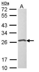
- Western Blot: Loricrin Antibody [NBP1-33610] - Sample (30 ug of whole cell lysate) A: Raji 12% SDS-PAGE, antibody at 1:1000.
- Submitted by
- Novus Biologicals (provider)
- Main image

- Experimental details
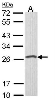
- Western Blot: Loricrin Antibody [NBP1-33610] - Whole cell extract (30 ug) was separated by 12% SDS-PAGE, and the membrane was blotted with Loricrin antibody [N1], N-term diluted at 1:1000. The HRP-conjugated anti-rabbit IgG antibody (NBP2-19301) was used to detect the primary antibody.
- Submitted by
- Novus Biologicals (provider)
- Main image
- Experimental details
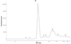
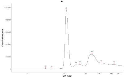
- Simple Western: Loricrin Antibody [NBP1-33610] - NHEK human normal epidermal keratinocytes. Antibody dilution of 1:50. Protein concentration of 450 ug/mL. Detection is chemiluminescence. Simple Western image submitted by a verified customer review.
Supportive validation
- Submitted by
- Novus Biologicals (provider)
- Main image
- Experimental details
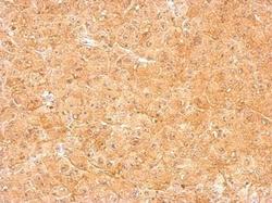
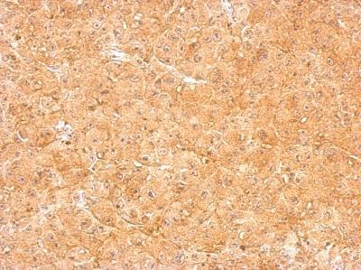
- Immunohistochemistry-Paraffin: Loricrin Antibody [NBP1-33610] - Paraffin-embedded HBL438 xenograft; antibody dilution 1:500.
- Submitted by
- Novus Biologicals (provider)
- Main image
- Experimental details
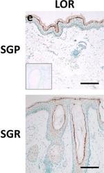
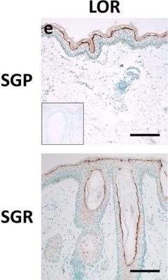
- Immunohistochemistry: Loricrin Antibody [NBP1-33610] - Prominent differences in the expressions of innate immune and barrier molecules between sebaceous gland poor (SGR) and sebaceous gland rich (SGP) skin regions. Representative images for immunostaining and quantification of epidermal levels of LOR in SGP and SGR skin sections. Images of negative control stainings are shown in the bottom left corner of SGP immunostainings. Size bars=100um. The graphs show the mean +/- SEM of measured protein levels (*p
- Submitted by
- Novus Biologicals (provider)
- Main image
- Experimental details
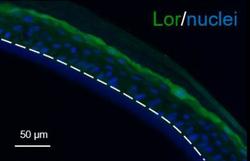
- Immunohistochemistry-Paraffin: Loricrin Antibody [NBP1-33610] - Reconstructed human epidermis tissues were embedded in paraffin. After successives baths in xylene and ethanol, antigen was retrieved using warm citrate (for 30 min). Specimens were incubated overnight with the primary antibody (1:100 dilution). Blue staining corresponds to nuclei while loricrin is visible in green. IHC-P image submitted by a verified customer review.
