 Explore
 Validate
 Learn
Learn Western blot
Antibody data
- Antibody Data
- Antigen structure
- References [0]
- Comments [0]
- Validations
- Western blot [2]
- Other assay [7]
Submit
Validation data
Reference
Comment
Report error
- Product number
- PA5-17495 - Provider product page

- Provider
- Invitrogen Antibodies
- Product name
- Phospho-MARK1/MARK2/MARK3 (Thr215, Thr208, Thr234) Polyclonal Antibody
- Antibody type
- Polyclonal
- Antigen
- Synthetic peptide
- Description
- This antibody does not react with MARK4. It is not recommended to aliquot this antibody.
- Reactivity
- Human, Mouse, Rat
- Host
- Rabbit
- Isotype
- IgG
- Vial size
- 100 µL
- Concentration
- 59 µg/mL
- Storage
- -20°C
No comments: Submit comment
Supportive validation
- Submitted by
- Invitrogen Antibodies (provider)
- Main image

- Experimental details
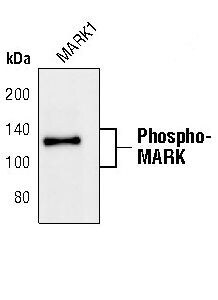
- Western blot analysis of Phospho-MARK Family in extracts from 293 cells transfected with either wild-type or threonine to alanine mutations at the respective phosphorylation sites of the MARK family members using Phospho-MARK Family polyclonal antibody (Product # PA5-17495). Transfected constructs contain a GST and HA tag. Transfection efficiency monitored with an HA antibody.
- Submitted by
- Invitrogen Antibodies (provider)
- Main image
- Experimental details
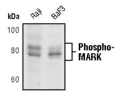
- Western blot analysis of Phospho-MARK Family in extracts from Raji and BaF3 cells showing endogenous levels of phosphorylated MARK family members using Phospho-MARK Family polyclonal antibody (Product # PA5-17495).

Supportive validation
- Submitted by
- Invitrogen Antibodies (provider)
- Main image
- Experimental details
- NULL
- Submitted by
- Invitrogen Antibodies (provider)
- Main image

- Experimental details
- NULL
- Submitted by
- Invitrogen Antibodies (provider)
- Main image

- Experimental details
- NULL
- Submitted by
- Invitrogen Antibodies (provider)
- Main image

- Experimental details
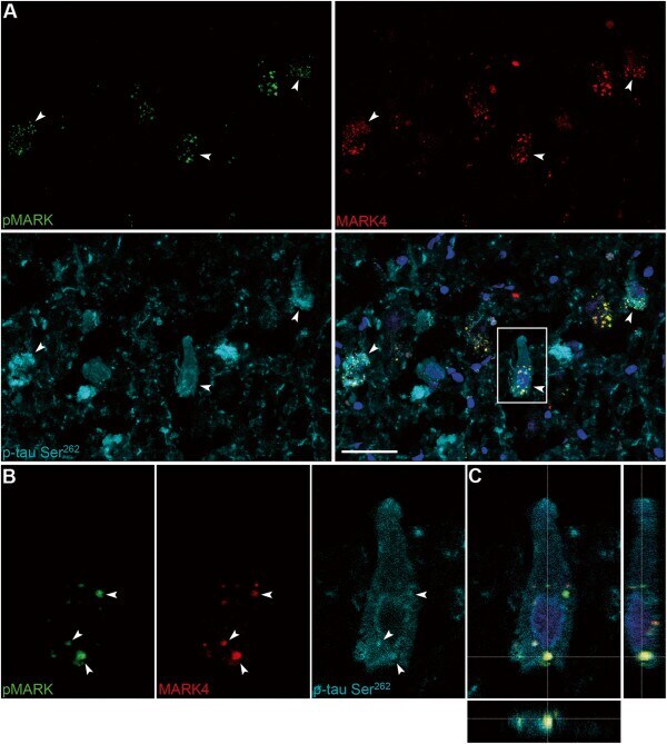
- Figure 8 Colocalization of MARK4, phospho-MARK, and p-tau Ser 262 in GVDs. Triple-immunofluorescence images from the CA1-field of an AD case (#32) with antibodies against pMARK (FITC), MARK4 (Cy3) and p-tau Ser 262 (Cy5). (A) Arrowheads indicate neurons that stain positively for all three antibodies. (B) A typical pre-tangle neuron with perinuclear phospho-tau staining is magnified, where arrowheads indicate individual GVDs where all three markers colocalize. (C) A 3-D reconstruction of a Z-stack focused on the GVD in the bottom part of the neuron. Scale bar: 100 mum.
- Submitted by
- Invitrogen Antibodies (provider)
- Main image
- Experimental details
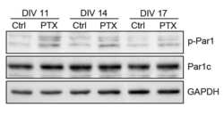
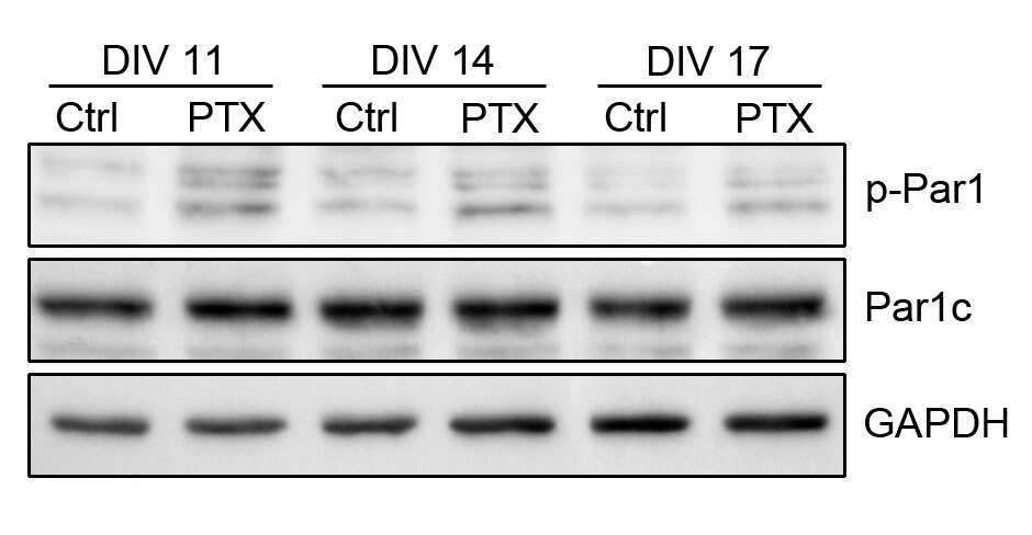
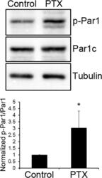
- Fig 1 MARK/Par1 is regulated by synaptic activity. Hippocampal neurons were treated with either DMSO (Control) or 10 muM of picrotoxin (PTX) for one hour. After treatment, neurons were lysed and immunoblotted with the indicated antibodies. Representative blots are shown in (a) and quantifications are shown in (b), expressed as Mean +- SD. *p

- Submitted by
- Invitrogen Antibodies (provider)
- Main image

- Experimental details
- Fig 2 MARK/Par1 is activated by NMDA receptors. a. Hippocampal neurons were pretreated with CNQX and TTX for 30 minutes, then stimulated with 50 muM of NMDA for 5 min. For inhibition of NMDA receptors, APV was included in the pretreatment and during NMDA stimulation. Neurons were lysed and immunoblotted with the indicated antibodies. b. Quantifications of blots in (a). Data are Mean +- SD, *p

- Submitted by
- Invitrogen Antibodies (provider)
- Main image
- Experimental details
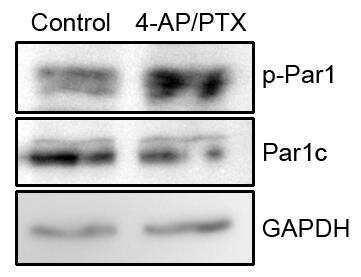
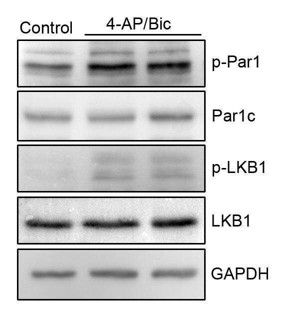
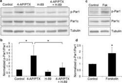
- Fig 3 MARK/Par1 is activated by NMDA receptors through PKA. a. Hippocampal neurons were treated with 4-AP and picrotoxin for 10 minutes. For inhibition of PKA, H-89 was included in the pretreatment and during 4-AP/PTX stimulation. Neurons were lysed and immunoblotted with the indicated antibodies. b. Quantifications of blots in (a). Data are Mean +- SD, *p
