Explore
 Validate
 Learn
Learn Western blot
Antibody data
- Antibody Data
- Antigen structure
- References [0]
- Comments [0]
- Validations
- Western blot [1]
- Immunohistochemistry [1]
Submit
Validation data
Reference
Comment
Report error
- Product number
- AF3435 - Provider product page

- Provider
- R&D Systems
- Product name
- Human/Mouse/Rat MLK4 alpha Antibody
- Antibody type
- Polyclonal
- Description
- Immunogen affinity purified. Detects endogenous human, mouse, and rat MLK4 alpha . Because the peptide immunogen corresponds to a region at the C-terminus of MLK4 alpha absent in MLK4 beta , no reactivity with MLK4 beta is present.
- Reactivity
- Human, Mouse, Rat
- Host
- Rabbit
- Conjugate
- Unconjugated
- Antigen sequence
CAC84639- Isotype
- IgG
- Vial size
- 100 ug
- Concentration
- LYOPH
- Storage
- Use a manual defrost freezer and avoid repeated freeze-thaw cycles. 12 months from date of receipt, -20 to -70 °C as supplied. 1 month, 2 to 8 °C under sterile conditions after reconstitution. 6 months, -20 to -70 °C under sterile conditions after reconstitution.
No comments: Submit comment
Supportive validation
- Submitted by
- R&D Systems (provider)
- Main image
- Experimental details
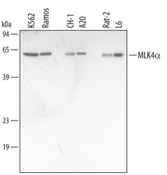
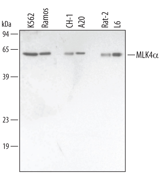
- Detection of Human/Mouse/Rat MLK4 alpha by Western Blot. Western blot shows lysates of K562 human chronic myelogenous leukemia cell line, Ramos human Burkitt's lymphoma cell line, A20 mouse B cell lymphoma cell line, CH-1 mouse B cell lymphoma cell line, Rat-2 rat embryonic fibroblast cell line, and L6 rat myoblast cell line. PVDF membrane was probed with 1 µg/mL of Human/Mouse/Rat MLK4 alpha Antigen Affinity-purified Polyclonal Antibody (Catalog # AF3435) followed by HRP-conjugated Anti-Rabbit IgG Secondary Antibody (Catalog # HAF008). A specific band was detected for MLK4 alpha at approximately 62 kDa (as indicated). This experiment was conducted under reducing conditions and using Immunoblot Buffer Group 1.
Supportive validation
- Submitted by
- R&D Systems (provider)
- Main image
- Experimental details
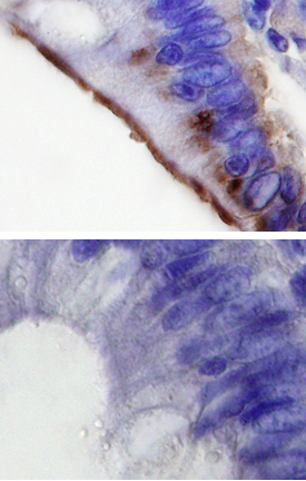
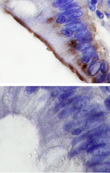
- MLK4 alpha in Human Colon Cancer Tissue. MLK4 alpha was detected in immersion fixed paraffin-embedded sections of human colon cancer tissue using 10 µg/mL Human/Mouse/Rat MLK4 alpha Antigen Affinity-purified Polyclonal Antibody (Catalog # AF3435) overnight at 4 °C. Before incubation with the primary antibody tissue was subjected to heat-induced epitope retrieval using Antigen Retrieval Reagent-Basic (Catalog # CTS013). Tissue was stained with the Anti-Rabbit HRP-DAB Cell & Tissue Staining Kit (brown; Catalog # CTS005) and counterstained with hematoxylin (blue). Specific labeling was localized to microvilli of epithelial cells. Lower panel shows a lack of labeling if primary antibodies are omitted and tissue is stained only with secondary antibody followed by incubation with detection reagents. View our protocol for Chromogenic IHC Staining of Paraffin-embedded Tissue Sections.