Explore
 Validate
 Learn
LearnPA5-27504
antibody from Invitrogen Antibodies
Targeting: ATP5F1A
ATP5A, ATP5A1, ATP5AL2, ATPM, hATP1, OMR, ORM
 Western blot
Antibody data
- Antibody Data
- Antigen structure
- References [1]
- Comments [0]
- Validations
- Western blot [6]
- Immunocytochemistry [2]
- Immunohistochemistry [5]
Submit
Validation data
Reference
Comment
Report error
- Product number
- PA5-27504 - Provider product page

- Provider
- Invitrogen Antibodies
- Product name
- ATP5A1 Polyclonal Antibody
- Antibody type
- Polyclonal
- Antigen
- Recombinant protein fragment
- Description
- Recommended positive controls: 293T, A431, HeLa, HepG2, K562, THP-1, mouse brain, rat brain. Predicted reactivity: Mouse (96%), Rat (96%), Zebrafish (90%), Xenopus laevis (89%), Dog (95%), Pig (96%), Chicken (90%), Rhesus Monkey (98%), Chimpanzee (100%), Bovine (96%). Store product as a concentrated solution. Centrifuge briefly prior to opening the vial.
- Reactivity
- Human, Mouse, Rat
- Host
- Rabbit
- Isotype
- IgG
- Vial size
- 100 µL
- Concentration
- 0.66 mg/mL
- Storage
- Store at 4°C short term. For long term storage, store at -20°C, avoiding freeze/thaw cycles.
Submitted references Nuclear cytoplasmic trafficking of proteins is a major response of human fibroblasts to oxidative stress.
Baqader NO, Radulovic M, Crawford M, Stoeber K, Godovac-Zimmermann J
Journal of proteome research 2014 Oct 3;13(10):4398-423
Journal of proteome research 2014 Oct 3;13(10):4398-423
No comments: Submit comment
Supportive validation
- Submitted by
- Invitrogen Antibodies (provider)
- Main image

- Experimental details
- Western Blot analysis of ATP5A1 was performed by separating 30 µg of non-transfected (–) and transfected (+) 293T whole cell extracts by 10% SDS-PAGE. Proteins were transferred to a membrane and probed with a ATP5A1 Polyclonal Antibody (Product # PA5-27504) at a dilution of 1:1000. The HRP-conjugated anti-rabbit IgG antibody was used to detect the primary antibody.

- Submitted by
- Invitrogen Antibodies (provider)
- Main image

- Experimental details
- Western Blot using ATP5A1 Polyclonal Antibody (Product # PA5-27504). HepG2 and mitochondria extracts (30 µg) were separated by SDS-PAGE, and the membrane was blotted with ATP5A1 Polyclonal Antibody (Product # PA5-27504) diluted at 1:2,000. The HRP-conjugated anti-rabbit IgG antibody was used to detect the primary antibody.

- Submitted by
- Invitrogen Antibodies (provider)
- Main image

- Experimental details
- Western Blot analysis of ATP5A1 was performed by separating 50 µg of various tissue extracts by 10% SDS-PAGE. Proteins were transferred to a membrane and probed with a ATP5A1 Polyclonal Antibody (Product # PA5-27504) at a dilution of 1:1,000.

- Submitted by
- Invitrogen Antibodies (provider)
- Main image

- Experimental details
- Western Blot analysis of ATP5A1 was performed by separating 30 µg of various whole cell extracts by 10% SDS-PAGE. Proteins were transferred to a membrane and probed with a ATP5A1 Polyclonal Antibody (Product # PA5-27504) at a dilution of 1:2000 and a HRP-conjugated anti-rabbit IgG secondary antibody.

- Submitted by
- Invitrogen Antibodies (provider)
- Main image

- Experimental details
- Western Blot analysis of ATP5A1 was performed by separating 30 µg of various whole cell extracts by 10% SDS-PAGE. Proteins were transferred to a membrane and probed with a ATP5A1 Polyclonal Antibody (Product # PA5-27504) at a dilution of 1:2,000.

- Submitted by
- Invitrogen Antibodies (provider)
- Main image
- Experimental details
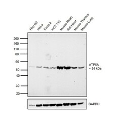
- Western blot was performed using Anti-ATP5A Rabbit Polyclonal Antibody (Product # PA5-27504) and a 54 kDa band corresponding to an isoform of ATP5A was observed in cell lines and tissues tested. Membrane enriched extracts (30 µg lysate) of Hep G2 (Lane 1), HeLa (Lane 2), Caco-2 (Lane 3), HCT 116 (Lane 4), Mouse Heart (Lane 5), Rat Heart (Lane 6), Mouse Thymus (Lane 7) and Mouse Lung (Lane 8) were electrophoresed using Novex® NuPAGE® 4-12% Bis-Tris Protein Gel (Product # NP0322BOX). Resolved proteins were then transferred onto a nitrocellulose membrane (Product # IB23001) by iBlot® 2 Dry Blotting System (Product # IB21001). The blot was probed with the primary antibody (1:1000 dilution) and detected by chemiluminescence with Goat anti-Rabbit IgG (H+L) Superclonal™ Recombinant Secondary Antibody, HRP (Product # A27036, 1:4000 dilution) using the iBright FL 1000 (Product # A32752). Chemiluminescent detection was performed using Novex® ECL Chemiluminescent Substrate Reagent Kit (Product # WP20005).

Supportive validation
- Submitted by
- Invitrogen Antibodies (provider)
- Main image

- Experimental details
- Immunocytochemistry-Immunofluorescence analysis of ATP5A1 was performed in HeLa cells fixed in 4% paraformaldehyde at RT for 15 min. Green: ATP5A1 Polyclonal Antibody (Product # PA5-27504) diluted at 1:500. Blue: Hoechst 33342 staining. Scale bar = 10 µm.

- Submitted by
- Invitrogen Antibodies (provider)
- Main image

- Experimental details
- Immunofluorescence analysis of ATP5A was performed using Hep G2 cells. The cells were fixed with 4% paraformaldehyde for 10 minutes, permeabilized with 0.1% Triton™ X-100 for 15 minutes, and blocked with 2% BSA for 1 hour at room temperature. The cells were labeled with mtTFA Rabbit Polyclonal Antibody (Product # PA5-27504) at 1:150 dilution in 0.1% BSA and incubated overnight at 4 degree and then labeled with Goat anti-Rabbit IgG (H+L) Highly Cross-Adsorbed Secondary Antibody, Alexa Fluor Plus 488 (Product # A32731) at a dilution of 1:2000 for 45 minutes at room temperature (Panel a: green). Nuclei (Panel b: blue) were stained with ProLong™ Diamond Antifade Mountant with DAPI (Product # P36962). F-actin (Panel c: red) was stained with Rhodamine Phalloidin (Product # R415, 1:300). Panel d represents the composite image showing mitochondrial pattern of ATP5A. Panel e represents control cells with no primary antibody to assess background. The images were captured at 60X magnification.

Supportive validation
- Submitted by
- Invitrogen Antibodies (provider)
- Main image

- Experimental details
- Immunohistochemistry (Frozen) analysis of ATP5A1 was performed in frozen sectioned adult mouse retina tissue using ATP5A1 Polyclonal Antibody (Product # PA5-27504) at a dilution of 1:250 (Green). Red: Protein kinase C alpha staining. Blue: Fluoroshield with DAPI.

- Submitted by
- Invitrogen Antibodies (provider)
- Main image
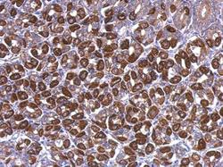
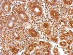
- Experimental details
- ATP5A1 Polyclonal Antibody detects ATP5A1 protein at cytoplasm on mouse stomach by immunohistochemical analysis. Sample: Paraffin-embedded mouse stomach. ATP5A1 Polyclonal Antibody (Product # PA5-27504) dilution: 1:500. Antigen Retrieval: EDTA based buffer, pH 8.0, 15 min.
- Submitted by
- Invitrogen Antibodies (provider)
- Main image
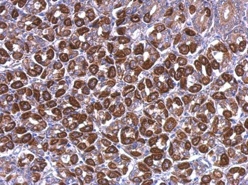
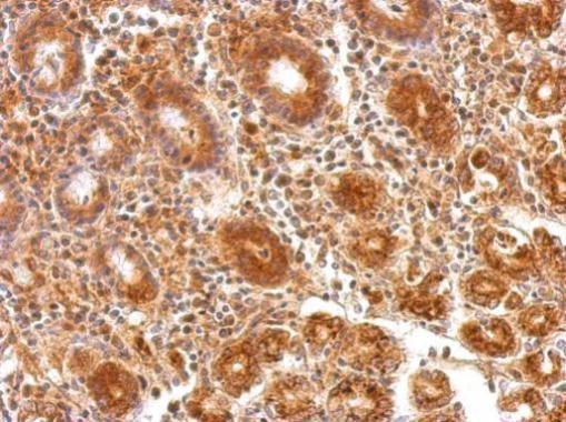
- Experimental details
- ATP5A1 Polyclonal Antibody detects ATP5A1 protein at cytosol on human colon carcinoma by immunohistochemical analysis. Sample: Paraffin-embedded colon carcinoma. ATP5A1 Polyclonal Antibody (Product # PA5-27504) dilution: 1:200. Antigen Retrieval: EDTA based buffer, pH 8.0, 15 min.
- Submitted by
- Invitrogen Antibodies (provider)
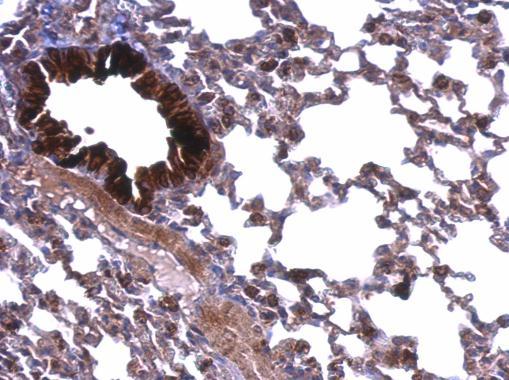
- Main image

- Experimental details
- ATP5A1 Polyclonal Antibody detects ATP5A1 protein at membrane on mouse lung by immunohistochemical analysis. Sample: Paraffin-embedded mouse lung. ATP5A1 Polyclonal Antibody (Product # PA5-27504) dilution: 1:500. Antigen Retrieval: EDTA based buffer, pH 8.0, 15 min.