Explore
 Validate
 Learn
LearnAF7005
antibody from R&D Systems
Targeting: VSIR
B7-H5, B7H5, C10orf54, Dies1, GI24, PD-1H, SISP1, VISTA
 Western blot
Antibody data
- Antibody Data
- Antigen structure
- References [0]
- Comments [0]
- Validations
- Western blot [1]
- Immunocytochemistry [1]
- Flow cytometry [1]
Submit
Validation data
Reference
Comment
Report error
- Product number
- AF7005 - Provider product page

- Provider
- R&D Systems
- Product name
- Mouse VISTA/B7-H5/PD-1H Antibody
- Antibody type
- Polyclonal
- Description
- Antigen Affinity-purified. Detects mouse VISTA/B7-H5/PD-1H in direct ELISAs and Western blots. In direct ELISAs, approximately 25% cross-reactivity with recombinant human VISTA/B7-H5/PD-1H is observed.
- Reactivity
- Mouse
- Host
- Sheep
- Conjugate
- Unconjugated
- Antigen sequence
Q9D659- Isotype
- IgG
- Vial size
- 100 ug
- Concentration
- LYOPH
- Storage
- Use a manual defrost freezer and avoid repeated freeze-thaw cycles. 12 months from date of receipt, -20 to -70 °C as supplied. 1 month, 2 to 8 °C under sterile conditions after reconstitution. 6 months, -20 to -70 °C under sterile conditions after reconstitution.
No comments: Submit comment
Supportive validation
- Submitted by
- R&D Systems (provider)
- Main image

- Experimental details
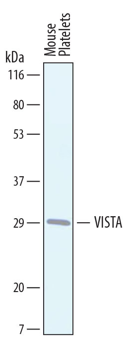
- Detection of VISTA/B7-H5/PD-1H by Western Blot. Western blot shows lysates of mouse platelets. PVDF membrane was probed with 1 µg/mL of Sheep Anti-Mouse VISTA/B7-H5/PD-1H Antigen Affinity-purified Polyclonal Antibody (Catalog # AF7005) followed by HRP-conjugated Anti-Sheep IgG Secondary Antibody (Catalog # HAF016). A specific band was detected for VISTA/B7-H5/PD-1H at approximately 30 kDa (as indicated). This experiment was conducted under reducing conditions and using Immunoblot Buffer Group 8.
Supportive validation
- Submitted by
- R&D Systems (provider)
- Main image

- Experimental details
- VISTA/B7-H5/PD-1H in D3 Mouse Cell Line. VISTA/B7-H5/PD-1H was detected in immersion fixed D3 mouse embryonic stem cell line using Sheep Anti-Mouse VISTA/B7-H5/PD-1H Antigen Affinity-purified Polyclonal Antibody (Catalog # AF7005) at 10 µg/mL for 3 hours at room temperature. Cells were stained using the NorthernLights™ 557-conjugated Anti-Sheep IgG Secondary Antibody (red; Catalog # NL010) and counterstained with DAPI (blue). Specific staining was localized to cell surfaces and cytoplasm. View our protocol for Fluorescent ICC Staining of Cells on Coverslips.

Supportive validation
- Submitted by
- R&D Systems (provider)
- Main image

- Experimental details
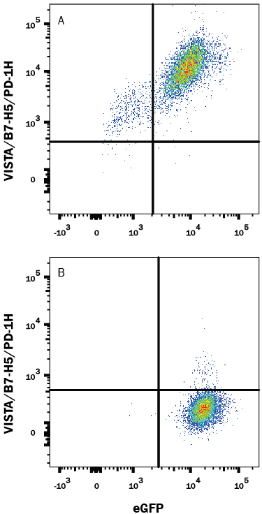
- Detection of VISTA/B7-H5/PD-1H in HEK293 Human Cell Line Transfected with Mouse VISTA and eGFP by Flow Cytometry. HEK293 human embryonic kidney cell line transfected with either (A) mouse VISTA or (B) irrelevant transfectants and eGFP was stained with Sheep Anti-Mouse VISTA/B7-H5/PD-1H Antigen Affinity-purified Polyclonal Antibody (Catalog # AF7005) followed by Allophycocyanin-conjugated Anti-Sheep IgG Secondary Antibody (Catalog # F0127). Quadrant markers were set based on control antibody staining (Catalog # 5-001-A). View our protocol for Staining Membrane-associated Proteins.