Explore
 Validate
 Learn
LearnAF7126-100
antibody from R&D Systems
Targeting: VSIR
B7-H5, B7H5, C10orf54, Dies1, GI24, PD-1H, SISP1, VISTA
 Western blot
Antibody data
- Antibody Data
- Antigen structure
- References [1]
- Comments [0]
- Validations
- Western blot [1]
- ELISA [1]
- Flow cytometry [1]
Submit
Validation data
Reference
Comment
Report error
- Product number
- AF7126-100 - Provider product page

- Provider
- R&D Systems
- Product name
- Human VISTA/B7-H5/PD-1H Antibody
- Antibody type
- Polyclonal
- Description
- Antigen Affinity-purified. Detects human VISTA/B7-H5/PD-1H in direct ELISAs and Western blots.
- Reactivity
- Human
- Host
- Sheep
- Conjugate
- Unconjugated
- Antigen sequence
Q9H7M9- Isotype
- IgG
- Vial size
- 100 ug
- Storage
- Use a manual defrost freezer and avoid repeated freeze-thaw cycles. 12 months from date of receipt, -20 to -70 °C as supplied. 1 month, 2 to 8 °C under sterile conditions after reconstitution. 6 months, -20 to -70 °C under sterile conditions after reconstitution.
Submitted references VSIG-3 as a ligand of VISTA inhibits human T-cell function.
Wang J, Wu G, Manick B, Hernandez V, Renelt M, Erickson C, Guan J, Singh R, Rollins S, Solorz A, Bi M, Li J, Grabowski D, Dirkx J, Tracy C, Stuart T, Ellinghuysen C, Desmond D, Foster C, Kalabokis V
Immunology 2019 Jan;156(1):74-85
Immunology 2019 Jan;156(1):74-85
No comments: Submit comment
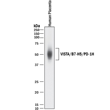
Supportive validation
- Submitted by
- R&D Systems (provider)
- Main image
- Experimental details
- Detection of Human VISTA/B7-H5/PD-1H by Western Blot. Western blot shows lysates of human placenta tissue. PVDF membrane was probed with 1 µg/mL of Sheep Anti-Human VISTA/B7-H5/PD-1H Antigen Affinity-purified Polyclonal Antibody (Catalog # AF7126) followed by HRP-conjugated Anti-Sheep IgG Secondary Antibody (Catalog # HAF016). A specific band was detected for VISTA/B7-H5/PD-1H at approximately 45-65 kDa (as indicated). This experiment was conducted under reducing conditions and using Immunoblot Buffer Group 1.
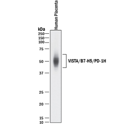
Supportive validation
- Submitted by
- R&D Systems (provider)
- Main image
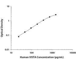
- Experimental details
- Human VISTA/B7-H5/PD-1H ELISA Standard Curve. Recombinant Human VISTA/B7-H5/PD-1H protein was serially diluted 2-fold and captured by Mouse Anti-Human VISTA/B7-H5/PD-1H Monoclonal Antibody (Catalog # MAB71264) coated on a Clear Polystyrene Microplate (Catalog # DY990). Sheep Anti-Human VISTA/B7-H5/PD-1H Antigen Affinity-purified Polyclonal Antibody (Catalog # AF7126) was biotinylated and incubated with the protein captured on the plate. Detection of the standard curve was achieved by incubating Streptavidin-HRP (Catalog # DY998) followed by Substrate Solution (Catalog # DY999) and stopping the enzymatic reaction with Stop Solution (Catalog # DY994).

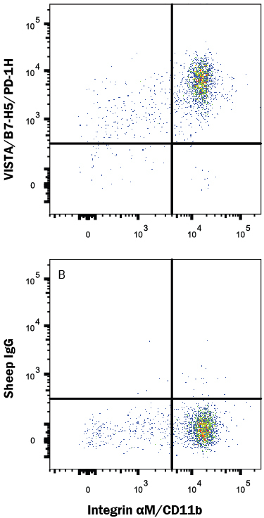
Supportive validation
- Submitted by
- R&D Systems (provider)
- Main image
- Experimental details
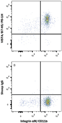
- Detection of VISTA/B7-H5/PD-1H in Human PBMCs by Flow Cytometry. Human peripheral blood mononuclear cells (PBMCs) were stained with Mouse Anti-Human CD11b/Integrin alpha M APC-conjugated Monoclonal Antibody (Catalog # FAB16991A) and either (A) Sheep Anti-Human VISTA/B7-H5/PD-1H Antigen Affinity-purified Polyclonal Antibody (Catalog # AF7126) or (B) Normal Sheep IgG Control (Catalog # 5-001-A) followed by Phycoerythrin-conjugated Anti-Sheep IgG Secondary Antibody (Catalog # F0126). View our protocol for Staining Membrane-associated Proteins.