 Explore
 Validate
 Learn
Learn Western blot
Antibody data
- Antibody Data
- Antigen structure
- References [0]
- Comments [0]
- Validations
- Western blot [1]
- ELISA [1]
- Other assay [3]
Submit
Validation data
Reference
Comment
Report error
- Product number
- TA347214 - Provider product page

- Provider
- OriGene
- Product name
- Rabbit Polyclonal H4K5ac Antibody
- Antibody type
- Polyclonal
- Description
- Rabbit Polyclonal H4K5ac Antibody
- Host
- Rabbit
- Conjugate
- Unconjugated
- Epitope
- HIST4H4
- Isotype
- IgG
- Antibody clone number
- NULL
- Vial size
- 50 µg
- Concentration
- 1.80 ?g/?l
No comments: Submit comment
Supportive validation
- Submitted by
- OriGene (provider)
- Main image

- Experimental details
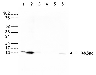
- WB was performed on whole cell (25 ug, lane 1) and histone extracts (15 ug, lane 2 ) from HeLa cells, and on 1 ug of recombinant histone H2A, H2B, H3 and H4 (lane 3, 4, 5 and 6, respectively) using the antibody against H4K5ac. The antibody was diluted 1:500 in TBS-Tween containing 5% skimmed milk. The marker (in kDa) is shown on the left.
- Validation comment
- WB
Supportive validation
- Submitted by
- OriGene (provider)
- Main image

- Experimental details
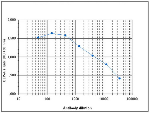
- Determination of the antibody titer To determine the titer of the antibody, an ELISA was performed using a serial dilution of the antibody against H4K5ac in antigen coated wells. The antigen used was a peptide containing the histone modification of interest. By plotting the absorbance against the antibody dilution (Figure 3), the titer of the antibody was estimated to be 1:8,900.
- Validation comment
- ELISA
Supportive validation
- Submitted by
- OriGene (provider)
- Main image

- Experimental details
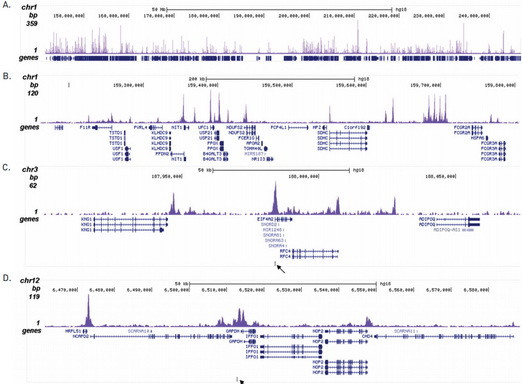
- ChIP was performed with 1 ug of the ab on sheared chromatin from 1 million HeLa cells. The 36 bp tags were aligned to the human genome using the ELAND algorithm. Image shows the signal distribution along the complete length of chromosome 1 and a zoomin to a 500 kb region (B). C and D show the enrichment in two genomic regions on chromosome 3 and 12, respectively, containing EIF4A2 and GAPDH positive controls. The position of the amplicon used for validating the QPCR results is shown with arrow.
- Validation comment
- Assay
- Submitted by
- OriGene (provider)
- Main image
- Experimental details
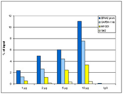
- ChIP assays using HeLa cells on sheared chromatin from 1 million cells. Titration of 1,2,5&10ug antibody per ChIP was analysed. IgG (2 ug/IP) was used as negative control. qPCR primers were for promoter of active gene EIF4A2 and for a region 1 kb upstream of GAPDH as positive controls, and for inactive MYOD1 gene and the Sat2 satellite repeat region as negative controls. Image shows the recovery, expressed as a % of input (the relative amount of IP'd DNA compared to input DNA after qPCR).
- Validation comment
- Assay

- Submitted by
- OriGene (provider)
- Main image

- Experimental details
- A Dot Blot analysis was performed with peptides containing other histone modifications and the unmodified H4. One hundred to 0.2 pmol of the respective peptides were spotted on a membrane. The antibody was used at a dilution of 1:5,000. Figure 4 shows a high specificity of the antibody for the modification of interest.
- Validation comment
- DB
