Explore
 Validate
 Learn
LearnPA5-19187
antibody from Invitrogen Antibodies
Targeting: CAVIN3
cavin-3, HSRBC, MGC20400, PRKCDBP, SRBC
 Western blot
Antibody data
- Antibody Data
- Antigen structure
- References [0]
- Comments [0]
- Validations
- Western blot [3]
- Immunohistochemistry [1]
Submit
Validation data
Reference
Comment
Report error
- Product number
- PA5-19187 - Provider product page

- Provider
- Invitrogen Antibodies
- Product name
- PRKCDBP Polyclonal Antibody
- Antibody type
- Polyclonal
- Antigen
- Synthetic peptide
- Description
- This antibody is predicted to react with bovine, canine and rat based on sequence homology. This antibody is tested in Peptide ELISA: antibody detection limit dilution 32,000.
- Reactivity
- Human, Mouse, Rat
- Host
- Goat
- Isotype
- IgG
- Vial size
- 100 µg
- Concentration
- 0.5 mg/mL
- Storage
- -20° C, Avoid Freeze/Thaw Cycles
No comments: Submit comment
Supportive validation
- Submitted by
- Invitrogen Antibodies (provider)
- Main image

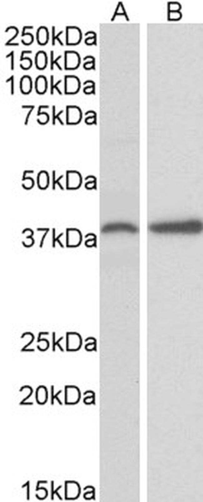
- Experimental details
- Western blot analysis of PRKCDBP in Mouse Ovary (A) and Rat Uterus (B) lysate (35µg protein in RIPA buffer). Samples were probed with the PRKCDBP antibody (Product # PA5-19187, 0.3µg/mL) for 1 hour. Western blot was detected by chemiluminescence.

- Submitted by
- Invitrogen Antibodies (provider)
- Main image

- Experimental details
- Western blot analysis of PRKCDBP by a PRKCDBP monoclonal antibody (Product # PA5-19187) at a concentration of 0.3 µg/mL. Mouse Ovary (A) and Rat Uterus (B) lysate (35µg protein in RIPA buffer). Detected by chemiluminescence.
- Submitted by
- Invitrogen Antibodies (provider)
- Main image
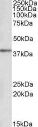
- Experimental details
- Western Blot staining of Human Adipose lysate using Product # PA5-19187 at a concentration of 0.1 µg/mL, the primary antibody incubation was 1 hour and the detection method was chemiluminescence.

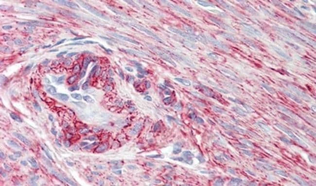
Supportive validation
- Submitted by
- Invitrogen Antibodies (provider)
- Main image

- Experimental details
- Immunohistochemical analysis of PRKCDBP in Human Uterus using a PRKCDBP monoclonal antibody (Product #PA5-19187) at 3.75 µg/mL. The Human Uterus tissue section was paraffin embeded and detected using steamed antigen retrieval with citrate buffer pH 6, AP-staining.