Explore
 Validate
 Learn
Learn Flow cytometry
Antibody data
- Antibody Data
- Antigen structure
- References [3]
- Comments [0]
- Validations
- Flow cytometry [1]
- Other assay [2]
Submit
Validation data
Reference
Comment
Report error
- Product number
- 11-0329-42 - Provider product page

- Provider
- Invitrogen Antibodies
- Product name
- CD32 Monoclonal Antibody (6C4 (CD32)), FITC, eBioscience™
- Antibody type
- Monoclonal
- Antigen
- Other
- Description
- Description: This 6C4 monoclonal antibody reacts with human CD32 (also known as Fc gammaRII). This antibody recognizes two isoforms of the receptor, Fc gammaRIIA and Fc gammaRIIB. These 40-kDa polymorphic transmembrane glycoproteins are expressed on B cells, granulocytes, monocytes, macrophages, and platelets. Moreover, these receptors are detected on natural killer cells. CD32 enables interaction between Fc gammaRII-expressing cells and opsonized antigen or IgG-containing immune complexes. Both receptors exhibit low affinity towards IgG and play a role in inflammation and autoimmune disease. This clone has been reported to inhibit Ig binding in a rosette blocking assay. Applications Reported: This 6C4 (CD32) antibody has been reported for use in flow cytometric analysis. Applications Tested: This 6C4 (CD32) antibody has been pre-titrated and tested by flow cytometric analysis of normal human peripheral blood cells. This can be used at 5 µL (0.25 µg) per test. A test is defined as the amount (µg) of antibody that will stain a cell sample in a final volume of 100 µL. Cell number should be determined empirically but can range from 10^5 to 10^8 cells/test. Excitation: 488 nm; Emission: 520 nm; Laser: Blue Laser. Filtration: 0.2 µm post-manufacturing filtered.
- Reactivity
- Human
- Host
- Mouse
- Conjugate
- Green dye
- Isotype
- IgG
- Antibody clone number
- 6C4 (CD32)
- Vial size
- 100 Tests
- Concentration
- 5 µL/Test
- Storage
- 4° C, store in dark, DO NOT FREEZE!
Submitted references Human Schwann cells are susceptible to infection with Zika and yellow fever viruses, but not dengue virus.
Immortalization of primary microglia: a new platform to study HIV regulation in the central nervous system.
Mechanisms controlling the effects of bevacizumab (avastin) on the inhibition of early but not late formed corneal neovascularization.
Dhiman G, Abraham R, Griffin DE
Scientific reports 2019 Jul 9;9(1):9951
Scientific reports 2019 Jul 9;9(1):9951
Immortalization of primary microglia: a new platform to study HIV regulation in the central nervous system.
Garcia-Mesa Y, Jay TR, Checkley MA, Luttge B, Dobrowolski C, Valadkhan S, Landreth GE, Karn J, Alvarez-Carbonell D
Journal of neurovirology 2017 Feb;23(1):47-66
Journal of neurovirology 2017 Feb;23(1):47-66
Mechanisms controlling the effects of bevacizumab (avastin) on the inhibition of early but not late formed corneal neovascularization.
Chen WL, Chen YM, Chu HS, Lin CT, Chow LP, Chen CT, Hu FR
PloS one 2014;9(4):e94205
PloS one 2014;9(4):e94205
No comments: Submit comment
Supportive validation
- Submitted by
- Invitrogen Antibodies (provider)
- Main image
- Experimental details
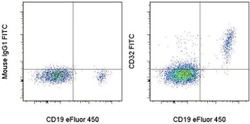
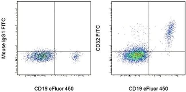
- Staining of normal human peripheral blood cells with Anti-Human CD19 eFluor® 450 (Product # 48-0199-42) and Mouse IgG1 K Isotype Control FITC (Product # 11-4714-42) (left) or Anti-Human CD32 FITC (right). Cells in the lymphocyte gate were used for analysis.
- Conjugate
- Green dye
Supportive validation
- Submitted by
- Invitrogen Antibodies (provider)
- Main image
- Experimental details
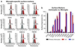
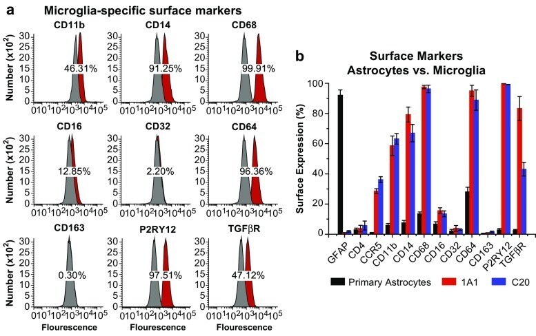
- Fig. 6 Surface expression of key markers of microglia. a Flow cytometry analysis was used to measure the surface expression of CD11b, CD14, CD68, CD16, CD32, CD64, CD163, P2RY12, and TGFbetaR on the representative immortalized cell line hmuglia 1A1. In each experiment, 100,000 cells were resuspended in 1 mL of cold PBS in the presence of 0.5 mug of the antibody or isotype control for 20 min on ice. Appropriate secondary antibodies were used in the absence of fluorophore-conjugated primary antibody, and the cell-antibody complexes were centrifuged and resuspended in PBS. In each FACS profile, the gray distributions represent the proportion of cells bound to the isotype control, whereas the red distributions represent the proportions of cells bound by the target antibody. b Quantification of the abovementioned markers as well as GFAP, CD4, and CCR5 on the surface of primary human astrocytes and immortalized microglia, as indicated
- Conjugate
- Green dye
- Submitted by
- Invitrogen Antibodies (provider)
- Main image
- Experimental details
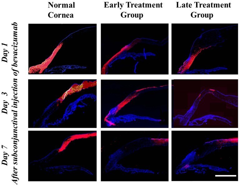
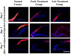
- Figure 1 Immunohistochemical staining of bevacizumab after subconjunctival injection as detected by a Cy3-conjugated IgG antibody. Time-dependent diffusion of bevacizumab into the corneal stroma was observed. Normal: eyes without induction of corneal NV. Bevacizumab molecule staining was observed in the entire thickness of corneal stroma adjacent to the injection site in all groups 1 day after the injection. The distribution of bevacizumab staining moved to the more central cornea on day 3 in all groups. On day 7, bevacizumab staining decreased dramatically in the peripheral cornea but was detected in the central cornea in all groups. Purple red: bevacizumab molecule staining; blue: DAPI staining of cell nuclei. Scale bar: 4 mm.
- Conjugate
- Green dye