Explore
 Validate
 Learn
Learn Western blot
Antibody data
- Antibody Data
- Antigen structure
- References [1]
- Comments [0]
- Validations
- Western blot [1]
- Immunocytochemistry [1]
- Other assay [1]
Submit
Validation data
Reference
Comment
Report error
- Product number
- PA5-66628 - Provider product page

- Provider
- Invitrogen Antibodies
- Product name
- FAM96A Polyclonal Antibody
- Antibody type
- Polyclonal
- Antigen
- Recombinant full-length protein
- Description
- Immunogen sequence: LATLIGLCLRV KLQRCLPFKH KLEIYISEGT HSTEEDINKQ INDKERVAAA Highest antigen sequence identity to the following orthologs - mouse 100%, rat 100%.
- Reactivity
- Human
- Host
- Rabbit
- Isotype
- IgG
- Vial size
- 100 µL
- Concentration
- 0.2 mg/mL
- Storage
- Store at 4°C short term. For long term storage, store at -20°C, avoiding freeze/thaw cycles.
Submitted references Viperin binds STING and enhances the type-I interferon response following dsDNA detection.
Crosse KM, Monson EA, Dumbrepatil AB, Smith M, Tseng YY, Van der Hoek KH, Revill PA, Saker S, Tscharke DC, G Marsh EN, Beard MR, Helbig KJ
Immunology and cell biology 2021 Apr;99(4):373-391
Immunology and cell biology 2021 Apr;99(4):373-391
No comments: Submit comment
Supportive validation
- Submitted by
- Invitrogen Antibodies (provider)
- Main image

- Experimental details
- Western blot analysis of FAM96A in human cell line RT-4, human cell line U-251 MG, human plasma, human liver tissue and human tonsil tissue. Samples were probed using a FAM96A Polyclonal Antibody (Product # PA5-66628).

Supportive validation
- Submitted by
- Invitrogen Antibodies (provider)
- Main image
- Experimental details
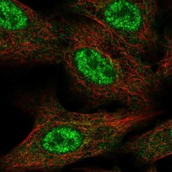
- Immunofluorescent staining of FAM96A in human cell line A549 shows localization to nucleoplasm and cytosol. Samples were probed using a FAM96A Polyclonal Antibody (Product # PA5-66628).

Supportive validation
- Submitted by
- Invitrogen Antibodies (provider)
- Main image
- Experimental details
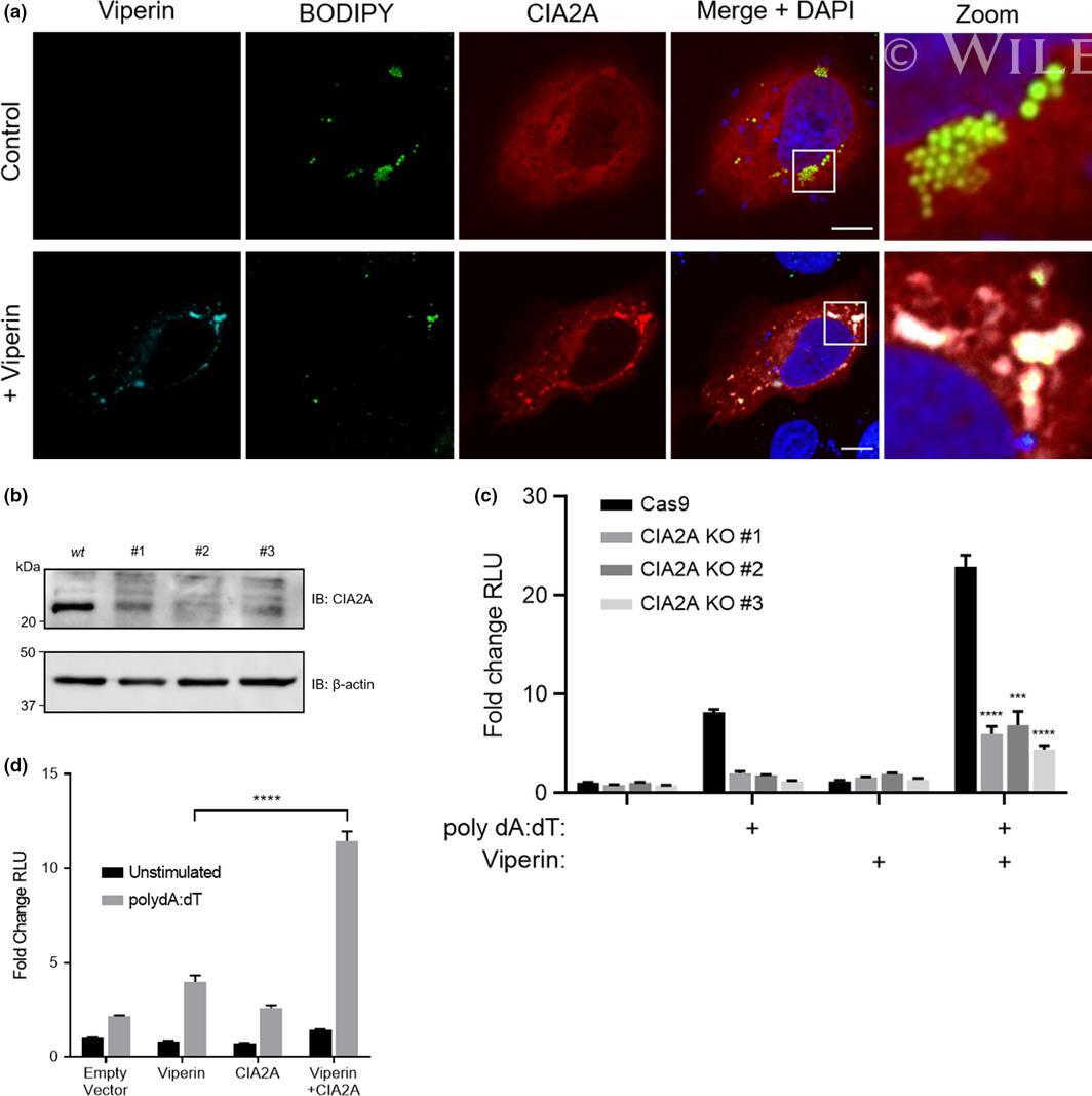
- 9 Figure Viperin co-localizes with CIA2A to enhance the type-I IFN response to dsDNA. (a) HeLa cells were transfected with viperin-flag and/or CIA2A-myc constructs 24 h prior to immunofluorescence staining with a rabbit monoclonal anti-flag antibody (Sigma) and/or mouse monoclonal anti-myc (Millipore), followed by an Alexa555-conjugated goat anti-rabbit (Invitrogen) and/or Alexa647-conjugated goat anti-mouse (Invitrogen) secondaries as well as BODIPY and DAPI staining. Imaged on a Ziess confocal LSM 780 microscope. The scale bar represents 10 um. Original magnification is x63. (b) Immunoblot analysis of CIA2A expression in HeLa wild-type (Cas9) and CRISPR-Cas9 polyclonal CIA2A knockdown cells; immunoblots are representative of two independent experiments. (c) Luciferase production driven by the IFN-beta promoter in HeLa wild-type (Cas9) and CRISPR-Cas9 polyclonal CIA2A knockdown cells transfected with viperin constructs 24 h prior to stimulation with poly dA: dT for 8 h. (d) Luciferase production driven by the IFN-beta promoter in HeLa cells transfected with viperin and/or CIA2A constructs 24 h prior to stimulation with poly dA: dT for 8 h. Luciferase measurements were controlled by constitutive expression of renilla and presented as fold changes in relative luminometer units (RLU) from Empty Vector unstimulated conditions. Equivalent results were obtained in at least three experiments. Data are presented as mean +- s.e.m. ; *** P < 0.001, **** P < 0.0001.