Explore
 Validate
 Learn
LearnPA5-20632
antibody from Invitrogen Antibodies
Targeting: MARCHF8
c-MIR, CMIR, MARCH-VIII, MARCH8, MIR, RNF178
 Western blot
Antibody data
- Antibody Data
- Antigen structure
- References [1]
- Comments [0]
- Validations
- Western blot [2]
- Immunocytochemistry [1]
- Immunohistochemistry [1]
- Other assay [5]
Submit
Validation data
Reference
Comment
Report error
- Product number
- PA5-20632 - Provider product page

- Provider
- Invitrogen Antibodies
- Product name
- MARCH8 Polyclonal Antibody
- Antibody type
- Polyclonal
- Antigen
- Synthetic peptide
- Description
- A suggested positive control is Hela cell lysate. PA5-20632 can be used with blocking peptide PEP-0752.
- Reactivity
- Human, Mouse, Rat
- Host
- Rabbit
- Isotype
- IgG
- Vial size
- 100 µg
- Concentration
- 1 mg/mL
- Storage
- Maintain refrigerated at 2-8°C for up to 3 months. For long term storage store at -20°C
Submitted references Elucidating the Antiviral Mechanism of Different MARCH Factors.
Umthong S, Lynch B, Timilsina U, Waxman B, Ivey EB, Stavrou S
mBio 2021 Mar 2;12(2)
mBio 2021 Mar 2;12(2)
No comments: Submit comment
Supportive validation
- Submitted by
- Invitrogen Antibodies (provider)
- Main image

- Experimental details
- Western blot analysis of HeLa cell lysate using a MARCH8 polyclonal antibody (Product # PA5-20632) at (A) 0.5 µg/mL and (B) 1 µg/mL.

- Submitted by
- Invitrogen Antibodies (provider)
- Main image

- Experimental details
- Western Blot analysis of MARCH8 in HeLa cell lysate with MARCH8 Polyclonal Antibody (Product # PA5-20632) at (A) 0.5 µg/mL and (B) 1 µg/mL.

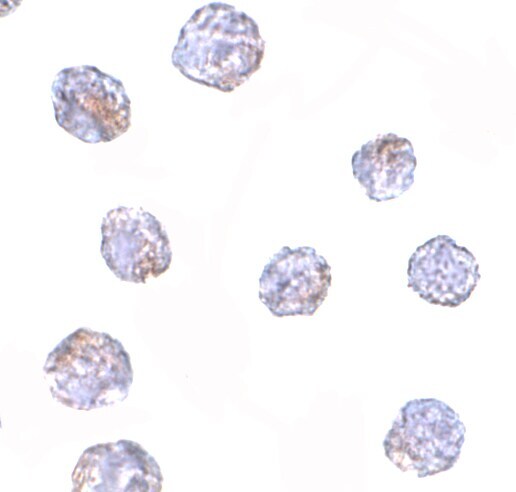
Supportive validation
- Submitted by
- Invitrogen Antibodies (provider)
- Main image

- Experimental details
- Immunocytochemistry of MARCH8 in HeLa cells with MARCH8 Polyclonal Antibody (Product # PA5-20632) at 2.5 µg/mL.
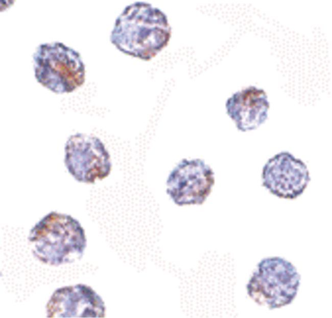
Supportive validation
- Submitted by
- Invitrogen Antibodies (provider)
- Main image

- Experimental details
- Immunocytochemistry staining of HeLa cells using a MARCH8 polyclonal antibody (Product # PA5-20632) at a 25 µg/mL dilution.
Supportive validation
- Submitted by
- Invitrogen Antibodies (provider)
- Main image
- Experimental details
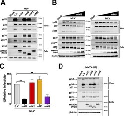
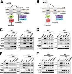
- FIG 2 Mouse March1 and mouse March8 block retrovirus infection by targeting the retroviral envelope glycoproteins. (A) Mouse March1 (mM1) and mM8 block the incorporation of the MLV envelope glycoproteins (p15E and gp70) in nascent virions by targeting them for degradation. (B) mM1 and mM8 target the MLV envelope glycoproteins for degradation in a dose-dependent manner. For both panels A and B, 293T cells were cotransfected with an MLV molecular clone and the indicated mouse March constructs. At 48 h posttransfection, cells and released virus in the culture medium were harvested, and the indicated proteins were analyzed by immunoblotting using anti-MLV p30 (detects both p30 and p65 Gag ), anti-MLV gp70, anti-MLV p15E/p12E, anti-myc (for detection of mM1, 2, 3, and 8), and anti-beta-actin antibodies. (C) Virus produced in the presence of mM1 and mM8 has decreased infectivity. NIH 3T3 cells were infected with equal amounts of 293T-derived MLV-luciferase reporter virus produced in the presence of mM1, 2, 3, or 8 or empty vector (E.V.). Cells were harvested 24 h postinfection, and luciferase levels were measured. The percentage (%) of relative infectivity was determined with respect to virus produced in the presence of E.V. All results are presented as means +- SD. Statistical significance was determined by one-way ANOVA. **, P < 0.01. (D) mM1 and mM8 target MMTV gp36 for degradation. 293T cells were cotransfected with an infectious genetically engineered MMTV hybrid provirus (HP)

- Submitted by
- Invitrogen Antibodies (provider)
- Main image
- Experimental details
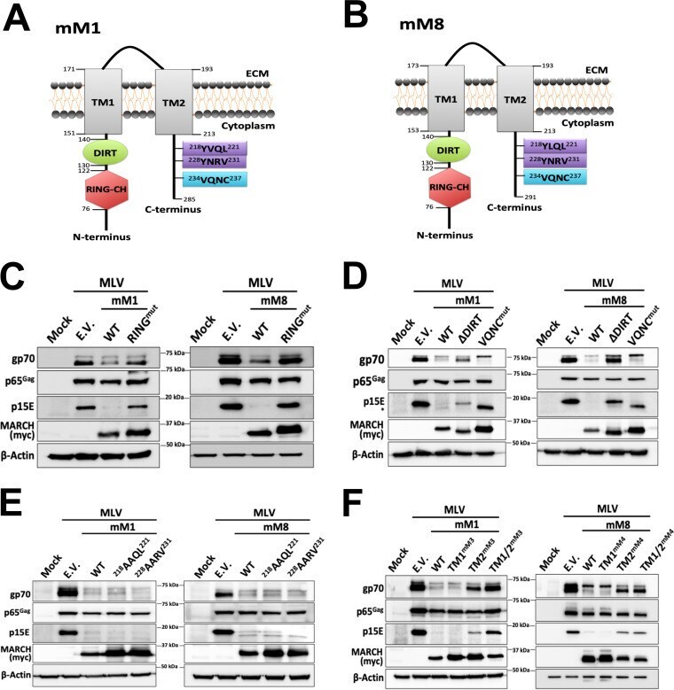
- FIG 4 The role of different mouse March1 and mouse March8 domains on retroviral envelope glycoprotein restriction. (A) Schematic diagram of mouse March (mM1) and (B) mM8 and their respective domains: Really Interesting New Gene-CH (RING-CH) domain, the d omain i n between R ING-CH domain and t ransmembrane domain (DIRT), N-terminal and C-terminal TM domains (TM1 and TM2, respectively), two tyrosine endocytic motifs, and a VQNC motif. (C to F) Only the RING-CH, DIRT, and TM2 domains of mM1 and 8 are important for the degradation of MLV envelope glycoprotein. 293T cells were cotransfected with either (C) RING-CH mutant (RING mut ) mM1 and mM8 , (D) mM1 and mM8 with the DIRT domain deleted (DeltaDIRT) or the VQNC domain mutated (VQNC mut ), (E) mM1 or mM8 with mutations in the tyrosine endocytic motifs and an MLV infectious clone, or (F) mM1 or mM8 with either the N-terminal (TM1), C-terminal (TM2), or both TM domains (TM1/2) swapped with those of mM3 (in the case of mM1 ) and mM4 (in the case of mM8 ). Cells were harvested 48 h posttransfection, and lysates were analyzed by immunoblotting using anti-MLV p30 (detects p65 Gag ), anti-MLV gp70, anti-MLV p15E, anti-myc (for detection of the mM1 and 8 proteins), and anti-beta-actin antibodies. Results are shown for n = 3 independent experiments. Representative immunoblotting results are shown for panels C through F.
- Submitted by
- Invitrogen Antibodies (provider)
- Main image
- Experimental details
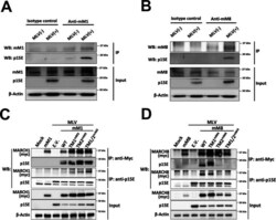
- FIG 6 Mouse March1 and mouse March8 physically interact with MLV p15E. (A and B) MutuDC1940 cells, which express endogenous levels of both mouse March1 (mM1) and mM8, were infected with MLV (10 MOI). Cells were harvested 72 h postinfection, and lysates were immunoprecipitated with (A and B) an isotype control, (A) anti-MARCH1, and (B) anti-MARCH8 and analyzed in Western blots (WB) with anti-p15E, anti-MARCH1, anti-MARCH8, and anti-beta-actin. (C and D) 293T cells were cotransfected with an MLV infectious clone and either (C) mouse March1 ( mM1 ) or (D) mM8 and their TM mutants used in Fig. 4 . Cells were harvested 48 h posttransfection, and lysates were immunoprecipitated with anti-myc (mM1 and mM8) and anti-p15E followed by Western blot analyses probing with anti-p15E, anti-myc (mM1 and mM8), and anti-beta-actin antibodies. Results are shown for n = 3 independent experiments. Representative immunoblotting results are shown for panels A through D.

- Submitted by
- Invitrogen Antibodies (provider)
- Main image
- Experimental details
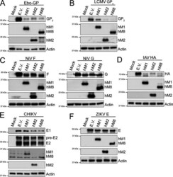
- FIG 9 MARCH proteins target envelope glycoproteins from a diverse number of viral families. (A) Ebola virus (EBOV) mature envelope glycoprotein Gp 2 , (B) lymphocytic choriomeningitis virus (LCMV) mature envelope glycoprotein GP 2 , (C) Nipah virus (NiV) fusion glycoprotein (F) (left) and NiV receptor binding glycoprotein (G) (right), (D) influenza A virus (IAV) hemagglutinin (HA), (E) Chikungunya virus (CHIKV) E1 and E2 protein, and (F) Zika virus (ZIKV) E levels in the presence of human MARCH1 (hM1), MARCH2 (hM2), MARCH8 (hM8), and empty vector (E.V). 293T cells were cotransfected with either (A) EBOV glycoprotein (Gp), (B) LCMV Gp, (C) NiV F or NiV G, (D) IAV HA, (E) a plasmid expressing the CHIKV structural proteins, or (F) ZIKV E in combination with either hM1 , 2 , 8 , or E.V. At 24 h posttransfection, cells were lysed and analyzed by Western blotting using anti-V5 (for EBOV GP 2 and IAV HA detection), anti-FLAG (for LCMV GP 2 detection), anti-AU1 (for NiV G detection), anti-HA (for NiV F detection), anti-flavivirus E antibody (4G2, for ZIKV E detection), anti-myc (CHIKV E1 detection), anti-E2 (CHIKV E2 detection), anti-MARCH1, anti-MARCH2, anti-MARCH8, and anti-beta-actin antibodies. Shown are the results of a single experiment (representative of three independent experiments).

- Submitted by
- Invitrogen Antibodies (provider)
- Main image
- Experimental details
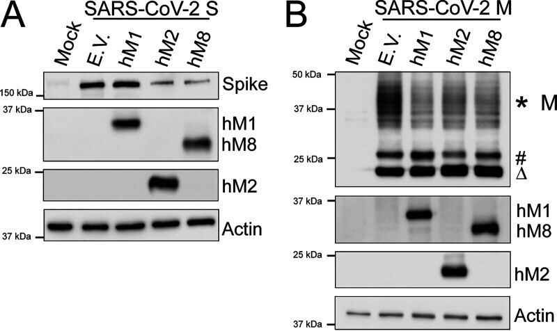
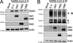
- FIG 10 MARCH proteins potently restrict the SARS-CoV-2 Spike (S) and Membrane (M) envelope glycoproteins. (A) SARS-CoV-2 Spike (S) and (B) SARS-CoV-2 Membrane (M) cellular levels in the presence of human MARCH1 (hM1), MARCH2 (hM2), MARCH8 (hM8), and empty vector (E.V.). 293T cells were cotransfected with SARS-CoV-2 S (A) or M (B) and either hM1 , 2 , 8 , or E.V. At 24 h posttransfection, cells were lysed and analyzed by Western blotting using anti-SARS-CoV-2 S, anti-V5 (for SARS-CoV-2 M detection), anti-MARCH1, anti-MARCH2, anti-MARCH8, and anti-beta-actin antibodies. The *, #, and Delta symbols represent different glycosylated forms of SARS-CoV-2 M. Shown are the results of a single experiment (representative of three independent experiments).