Explore
 Validate
 Learn
Learn Western blot
Antibody data
- Antibody Data
- Antigen structure
- References [0]
- Comments [0]
- Validations
- Western blot [1]
- Immunocytochemistry [2]
- Immunoprecipitation [1]
Submit
Validation data
Reference
Comment
Report error
- Product number
- ASM23 - Provider product page

- Provider
- Cytoskeleton, Inc.
- Product name
- Anti-SUMO-2/3
- Antibody type
- Monoclonal
- Antigen
- Full-length recombinant SUMO-2 protein (Uniprot: P61956) combined with a proprietary mix of peptides that include CQIRFRFDGQPINE.
- Description
- Anti-SUMO-2/3 mouse monoclonal antibody was raised against full-length recombinant SUMO-2 protein (Uniprot: P61956) combined with a proprietary mix of peptides that include CQIRFRFDGQPINE. The antibody has been shown to recognize a wide range of SUMO-2/3-targeted proteins in HeLa cell lysate (Fig. 1B) and to detect sub-nanogram amounts of recombinant SUMO-2 (Fig. 1A). Epitope mapping has identified that the antibody recognizes a sequence/structure within the peptide CQIRFRFDGQPINE. The peptide sequence is conserved in mammals, birds, and amphibians, giving the antibody broad species reactivity. ASM23 is purified by Protein G affinity chromatography and is supplied as a lyophilized white powder. Each Lot of antibody is quality controlled to provide a high batch to batch consistency. The Lot specific µg per tube can be found in the Lot specific COA documents at www.cytoskeleton.com.
- Host
- Mouse
- Conjugate
- Unconjugated
- Epitope
- CQIRFRFDGQPINE
- Isotype
- IgG
- Antibody clone number
- 12F3
- Vial size
- 2 x 100 µl
- Concentration
- Specified in COA
- Storage
- Lyophilized 4 degrees C. Reconstituted -20 degrees C.
- Handling
- Shipped at room temperture. The lyophilized protein can be stored desiccated at 4 degrees C for 6 months. Store at -20 degrees C when reconstituted.
No comments: Submit comment
Supportive validation
- Submitted by
- Cytoskeleton, Inc. (provider)
- Main image
- Experimental details
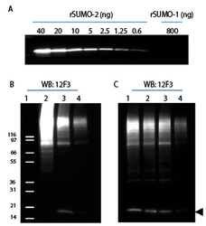
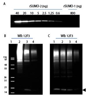
- 12F3 was used for immuno-blotting (1:500 dilution) following the recommended Western blot protocol (see below). Figure 1A: Titrations of recombinant SUMO-2 (40-0.6 ng) and SUMO-1 (800 ng). SUMO-2 was detected down to 0.6 ng while SUMO-1 was not detected at 800 ng. Figure 1B: Induction of SUMOylation by heat shock and reduction of SUMOylation by SUMO-2 shRNA knockdown. Cell lysates were prepared from HeLa cells: Lane 2: Heat Shock treated (43°C for 10min), Lane 3: untreated, Lane 4: shRNA SUMO-2 knock down. 20µg of HeLa cell lysates were used for each sample. Lane 1: position of molecular weight markers. Figure 1C: Specificity of SUMO-2 knockdown signal. Lane 1: parental HeLa cell lysates, Lane 2: SUMO-2 shRNA control lysates, Lane 3: SUMO-1 shRNA knock-down cell lysates, Lane 4: SUMO-2 shRNA knock-down cell lysates. Arrow head indicates free SUMO-2/3. To see the full Western blot protocol, see the product datasheet.
- Sample type
- Cell lysates were prepared from HeLa cells
- Primary Ab dilution
- 1:500
- Secondary Ab
- goat anti mouse secondary antibody
- Secondary Ab dilution
- 1:20,000
- Protocol
- Protocol
Supportive validation
- Submitted by
- Cytoskeleton, Inc. (provider)
- Main image
- Experimental details
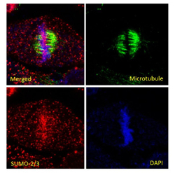
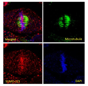
- HeLa cells were stained and visualized by confocal fluorescence microscopy as described in the IF method below. The cells were stained against ?/?-tubulin (sheep anti-tubulin Ab, Cat# ATN02, green) and SUMO-2/3 (12F3, red). DNA was stained with DAPI. Mitotic cells in metaphase were imaged with a Zeiss LSM 780 confocal microscope (1.4 NA 63X objective). Enrichment of SUMO 2/3 at chromosomes can be observed during mitosis as has been previously reported10. To see the full Immunofluorescence protocol, see the product datasheet.
- Sample type
- HeLa cells
- Primary Ab dilution
- 1:500
- Secondary Ab
- fluorescently-labeled donky anti-mouse
- Secondary Ab dilution
- 1:500
- Protocol
- Protocol
- Submitted by
- Cytoskeleton, Inc. (provider)
- Main image
- Experimental details
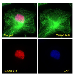
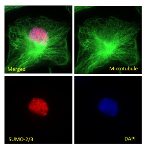
- HeLa cells were stained and visualized by widefield fluorescence microscopy as described in the IF method below. The cells were stained against ?/?-tubulin (sheep anti-tubulin Ab, Cat# ATN02, green) and SUMO-2/3 (12F3, red). DNA was stained with DAPI. Cells in interphase were imaged with a Zeiss Axio Observer.Z1 microscope (1.4 NA 63X objective). PML nuclear bodies (nuclear dots) were visible in SUMO-2/3 staining as has been previously reported11. To see the full Immunofluorescence protocol, see the product datasheet.
- Sample type
- HeLa cells
- Primary Ab dilution
- 1:500
- Secondary Ab
- fluorescently-labeled donky anti-mouse
- Secondary Ab dilution
- 1:500
- Protocol
- Protocol
Supportive validation
- Submitted by
- Cytoskeleton, Inc. (provider)
- Main image
- Experimental details
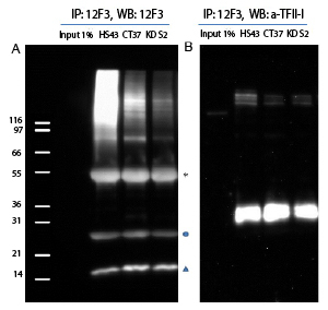
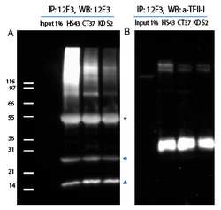
- Denatured cell lysates were prepared from HS43, CT37 and KD S212. 1mg of lysate was used for the immunoprecipitation of SUMO-2/3 conjugates. IP experiments were performed by the protocol presented in IP Method. Western blots of immunoprecipitated proteins were developed using 12F3 (A) or anti-TFII-I antibody (B). (A) Star (*) and circle (o) indicate heavy and light chains of antibodies. Unconjugated free SUMO is denoted by triangle. (B) Unconjugated TFII-I is visible near 120kDa. Multiple bands indicate that TFII-I is SUMOylated by several SUMO-2/3 proteins. TFII-I has previously been reported to be a target for Sumoylation . To see the full Immunoprecipitation protocol, see the product datasheet.
- Sample type
- Denatured cell lysates were prepared from HS43, CT37 and KD S2
- Primary Ab dilution
- Assay Dependent
- Secondary Ab
- Secondary Ab
- Protocol
- Protocol