 Explore
 Validate
 Learn
Learn Western blot
Antibody data
- Antibody Data
- Antigen structure
- References [0]
- Comments [0]
- Validations
- Western blot [2]
- Immunohistochemistry [4]
Submit
Validation data
Reference
Comment
Report error
- Product number
- LS-C290487 - Provider product page

- Provider
- LSBio
- Product name
- H2AFX / H2AX Antibody (C-Terminus) LS-C290487
- Antibody type
- Polyclonal
- Description
- Affinity purified
- Reactivity
- Human, Mouse
- Host
- Goat
- Isotype
- IgG
- Storage
- Store at 2-8°C for up to one year.
No comments: Submit comment
Supportive validation
- Submitted by
- LSBio (provider)
- Enhanced method
- Genetic validation
- Main image

- Experimental details
- Detection of Human H2AX by Western Blot. Samples: Whole cell lysate from 293T (15 and 50 ug) cells. Antibodies: Affinity purified goat anti-H2AX antibody used for WB at 0.4 ug/ml. Detection: Chemiluminescence with an exposure time of 30 seconds.

- Submitted by
- LSBio (provider)
- Enhanced method
- Genetic validation
- Main image

- Experimental details
- Detection of human H2AX by western blot. Samples: Whole cell lysate (50 µg) from HeLa, HEK293T, Jurkat, and K-562cells prepared using RIPA lysis buffer. Antibody: Affinity purified rabbit anti-H2AX antibody used for WB at 0.1 µg/ml. Detection: Chemiluminescence with an exposure time of 30 seconds.

Supportive validation
- Submitted by
- LSBio (provider)
- Enhanced method
- Genetic validation
- Main image

- Experimental details
- Detection of Mouse H2AX by Immunohistochemistry. Sample: FFPE section of mouse renal cell carcinoma. Antibody: Affinity purified rabbit anti-H2AX used at a dilution of 1:1000 (1 ug/ml). Detection: DAB.
- Submitted by
- LSBio (provider)
- Enhanced method
- Genetic validation
- Main image

- Experimental details
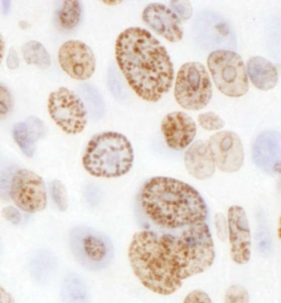
- Detection of Human H2AX by Immunohistochemistry. Sample: FFPE section of human lung cancer. Antibody: Affinity purified rabbit anti-H2AX used at a dilution of 1:5000 (0.2 ug/ml). Detection: DAB.

- Submitted by
- LSBio (provider)
- Enhanced method
- Genetic validation
- Main image

- Experimental details
- Detection of mouse H2AX by immunohistochemistry. Sample: FFPE section of mouse CT26 colon carcinoma. Antibody: Affinity purified goat anti- H2AX used at a dilution of 1:1,000 (1µg/ml). Detection: DAB

- Submitted by
- LSBio (provider)
- Enhanced method
- Genetic validation
- Main image

- Experimental details
- Detection of human H2AX by immunohistochemistry. Sample: FFPE section of human ovarian cancer. Antibody: Affinity purified goat anti- H2AX used at a dilution of 1:5,000 (0.2µg/ml). Detection: DAB
