Explore
 Validate
 Learn
Learn Flow cytometry
Antibody data
- Antibody Data
- Antigen structure
- References [2]
- Comments [0]
- Validations
- Flow cytometry [2]
- Other assay [2]
Submit
Validation data
Reference
Comment
Report error
- Product number
- 46-9865-42 - Provider product page

- Provider
- Invitrogen Antibodies
- Product name
- Phospho-Histone H2A.X (Ser139) Monoclonal Antibody (CR55T33), PerCP-eFluor™ 710, eBioscience™
- Antibody type
- Monoclonal
- Antigen
- Other
- Description
- Description: The CR55T33 monoclonal antibody recognizes phosphorylated serine 139 of human and mouse H2AX. H2AX is a member of the H2A histone family that complex with DNA and other histones to form the repeating nucleosome units characteristic of eukaryotic chromatin. Nucleosomes consist of approximately 147 base pairs of DNA wrapped around an octamer of histones composed of two each of the four histone proteins: H2A, H2B, H3 and H4. After induction of DNA damage such as double-strand breaks by irradiation, genotoxic stresses, replication errors or gene recombination, PI3K-like kinases (e.g., ataxia telangiectasia mutated (ATM), ataxia telangiectasia Rad-3-related (ATR), and DNA-dependent protein kinase (DNA-PK) are activated to phosphorylate serine 139 in H2AX. This early phosphorylation event plays a critical role in recruiting proteins involved in DNA repair. The monoclonal antibody CR55T33 recognizes a single band of approximately 15 kDa on reduced cell lysates from Jurkat cells stimulated with etoposide. Applications Reported: This CR55T33 antibody has been reported for use in intracellular staining followed by flow cytometric analysis. Applications Tested: This CR55T33 antibody has been pre-titrated and tested by intracellular staining followed by flow cytometric analysis of treated human peripheral blood cells using the Foxp3/Transcription Factor Buffer Set (Product # 00-5523-00) and protocol . Refer to BestProtocols®; Staining intracellular Antigens for Flow Cytometry Protocol (Protocol B: One step protocol for (nuclear) intracellular proteins). This can be used at 5 µL (0.125 µg) per test. A test is defined as the amount (µg) of antibody that will stain a cell sample in a final volume of 100 µL. Cell number should be determined empirically but can range from 10^5 to 10^8 cells/test. PerCP-eFluor® 710 emits at 710 nm and is excited with the blue laser (488 nm); it can be used in place of PerCP-Cyanine5.5. We recommend using a 710/50 bandpass filter, however, the 695/40 bandpass filter is an acceptable alternative. Please make sure that your instrument is capable of detecting this fluorochrome. Fixation: Samples can be stored in IC Fixation Buffer (Product # 00-8222) (100 µL of cell sample + 100 µL of IC Fixation Buffer) or 1-step Fix/Lyse Solution (Product # 00-5333) for up to 3 days in the dark at 4°C with minimal impact on brightness and FRET efficiency/compensation. Some generalizations regarding fluorophore performance after fixation can be made, but clone specific performance should be determined empirically. Excitation: 488 nm; Emission: 710 nm; Laser: Blue Laser. Filtration: 0.2 µm post-manufacturing filtered.
- Reactivity
- Human, Mouse
- Host
- Mouse
- Isotype
- IgG
- Antibody clone number
- CR55T33
- Vial size
- 100 Tests
- Concentration
- 5 μL/Test
- Storage
- 4°C, store in dark, DO NOT FREEZE!
Submitted references The mitotic checkpoint is a targetable vulnerability of carboplatin-resistant triple negative breast cancers.
ATR maintains chromosomal integrity during postnatal cerebellar neurogenesis and is required for medulloblastoma formation.
Moens S, Zhao P, Baietti MF, Marinelli O, Van Haver D, Impens F, Floris G, Marangoni E, Neven P, Annibali D, Sablina AA, Amant F
Scientific reports 2021 Feb 4;11(1):3176
Scientific reports 2021 Feb 4;11(1):3176
ATR maintains chromosomal integrity during postnatal cerebellar neurogenesis and is required for medulloblastoma formation.
Lang PY, Nanjangud GJ, Sokolsky-Papkov M, Shaw C, Hwang D, Parker JS, Kabanov AV, Gershon TR
Development (Cambridge, England) 2016 Nov 1;143(21):4038-4052
Development (Cambridge, England) 2016 Nov 1;143(21):4038-4052
No comments: Submit comment
Supportive validation
- Submitted by
- Invitrogen Antibodies (provider)
- Main image

- Experimental details
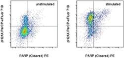
- Intracellular staining of 3-day unstimulated (left) or 600 uM etoposide-treated (right) normal humanperipheral blood cells with Anti-Human PARP1 (Cleaved) PE (Product # 12-6668-42) and Anti-Human/Mouse phospho-H2AX (S139) PerCP-eFluor® 710 using the Foxp3/Transcription Factor Staining Buffer Set (Product # 00-5523-00) and protocol. Cells in the lymphocyte gate were used for analysis.

- Submitted by
- Invitrogen Antibodies (provider)
- Main image
- Experimental details
- Intracellular staining of 3-day unstimulated (left) or 600 uM etoposide-treated (right) normal humanperipheral blood cells with Anti-Human PARP1 (Cleaved) PE (Product # 12-6668-42) and Anti-Human/Mouse phospho-H2AX (S139) PerCP-eFluor® 710 using the Foxp3/Transcription Factor Staining Buffer Set (Product # 00-5523-00) and protocol. Cells in the lymphocyte gate were used for analysis.

Supportive validation
- Submitted by
- Invitrogen Antibodies (provider)
- Main image

- Experimental details
- NULL

- Submitted by
- Invitrogen Antibodies (provider)
- Main image
- Experimental details
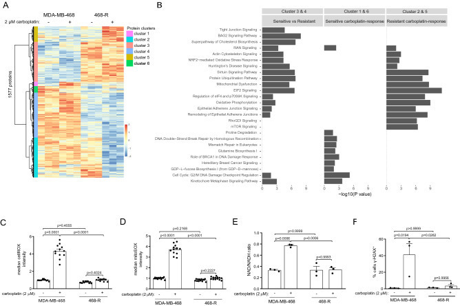
- Proteome re-wiring of carboplatin-resistant 468-R cells protects from carboplatin-induced oxidative and genotoxic stress. ( A,B ) Hierarchical clustering of differentially expressed proteins (DE) ( A ) and Ingenuity Pathway Analysis ( B ) of the proteome alterations in MDA-MB-468 and 468-R cells treated with vehicle or 2 uM carboplatin for 5 days. Protein expression was analyzed by LC/MS-MS, and DE proteins were identified by ANOVA followed by Tukey's HSD test with an adjusted P value < 0.05. The top 10 significantly enriched pathways are shown. P value < 0.05 was used to determine significantly enriched pathways; the -log10 ( P value) was not shown if the pathway was not significantly enriched. N = 3. ( C,D ) ROS levels in MDA-MB-468 and 468-R cells treated with vehicle or 2 uM carboplatin for 5 days. Total ROS levels ( C ) were measured by the cellROX assay, and mitochondrial specific superoxide levels ( D ) were measured by the mitoSOX assay. Median flow of cytometric intensities were normalized to MDA-MB-468 vehicle condition. Data are shown as Mean +- SEM. P values were calculated by two-way ANOVA with Geisser-Greenhouse and Tukey's correction. N = 3. ( E ) NAD/NADH ratio as measured by NAD/NADH-Glo kit. Data are shown as Mean +- SEM. P values were calculated by two-way ANOVA with Geisser-Greenhouse and Tukey's correction. N = 3. ( F ) gamma-H2AX staining of MDA-MB-468 and 468-R cells treated with vehicle or 2 uM carboplatin for 5 days as analyzed by flow cytometry. P va