Explore
 Validate
 Learn
Learn Immunohistochemistry
Antibody data
- Antibody Data
- Antigen structure
- References [3]
- Comments [0]
- Validations
- Immunohistochemistry [3]
- Other assay [3]
Submit
Validation data
Reference
Comment
Report error
- Product number
- 53-9865-82 - Provider product page

- Provider
- Invitrogen Antibodies
- Product name
- Phospho-Histone H2A.X (Ser139) Monoclonal Antibody (CR55T33), Alexa Fluor™ 488, eBioscience™
- Antibody type
- Monoclonal
- Antigen
- Other
- Description
- Description: The CR55T33 monoclonal antibody recognizes phosphorylated serine 139 of human and mouse H2AX. H2AX is a member of the H2A histone family that complex with DNA and other histones to form the repeating nucleosome units characteristic of eukaryotic chromatin. Nucleosomes consist of approximately 147 base pairs of DNA wrapped around an octamer of histones composed of two each of the four histone proteins: H2A, H2B, H3 and H4. After induction of DNA damage such as double-strand breaks by irradiation, genotoxic stresses, replication errors or gene recombination, PI3K-like kinases (e.g., ataxia telangiectasia mutated (ATM), ataxia telangiectasia Rad-3-related (ATR), and DNA-dependent protein kinase (DNA-PK) are activated to phosphorylate serine 139 in H2AX. This early phosphorylation event plays a critical role in recruiting proteins involved in DNA repair.
- Conjugate
- Green dye
- Antibody clone number
- CR55T33
- Concentration
- 0.5 mg/mL
Submitted references NCOA4 links iron bioavailability to DNA metabolism.
Synergistic lethality between PARP-trapping and alantolactone-induced oxidative DNA damage in homologous recombination-proficient cancer cells.
ATR maintains chromosomal integrity during postnatal cerebellar neurogenesis and is required for medulloblastoma formation.
Federico G, Carrillo F, Dapporto F, Chiariello M, Santoro M, Bellelli R, Carlomagno F
Cell reports 2022 Aug 16;40(7):111207
Cell reports 2022 Aug 16;40(7):111207
Synergistic lethality between PARP-trapping and alantolactone-induced oxidative DNA damage in homologous recombination-proficient cancer cells.
Wang H, Zhang S, Song L, Qu M, Zou Z
Oncogene 2020 Apr;39(14):2905-2920
Oncogene 2020 Apr;39(14):2905-2920
ATR maintains chromosomal integrity during postnatal cerebellar neurogenesis and is required for medulloblastoma formation.
Lang PY, Nanjangud GJ, Sokolsky-Papkov M, Shaw C, Hwang D, Parker JS, Kabanov AV, Gershon TR
Development (Cambridge, England) 2016 Nov 1;143(21):4038-4052
Development (Cambridge, England) 2016 Nov 1;143(21):4038-4052
No comments: Submit comment
Supportive validation
- Submitted by
- Invitrogen Antibodies (provider)
- Main image
- Experimental details
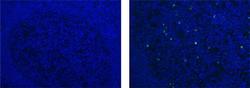
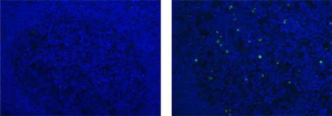
- Immunohistochemistry of formalin-fixed paraffin embedded human tonsil using 10 µg/mL of Mouse IgG1 K Isotype Control Alexa Fluor® 488 (left) or 10 µg/mL of Anti-Human/Mouse phospho-H2AX (S139) Alexa Fluor® 488 (right). Nuclei are stained with DAPI.
- Conjugate
- Green dye
- Submitted by
- Invitrogen Antibodies (provider)
- Main image

- Experimental details
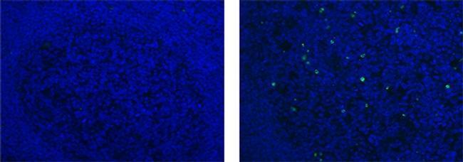
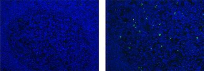
- Immunohistochemistry of formalin-fixed paraffin embedded human tonsil using 10 µg/mL of Mouse IgG1 K Isotype Control Alexa Fluor® 488 (left) or 10 µg/mL of Anti-Human/Mouse phospho-H2AX (S139) Alexa Fluor® 488 (right). Nuclei are stained with DAPI.
- Conjugate
- Green dye
Supportive validation
- Submitted by
- Invitrogen Antibodies (provider)
- Main image

- Experimental details
- NULL
- Conjugate
- Green dye

- Submitted by
- Invitrogen Antibodies (provider)
- Main image
- Experimental details
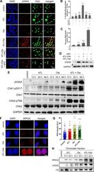
- Fig. 4 ATL synergizes with olaparib to induce intense replication stress in cancer cells. a Immunofluorescent staining of gammaH2AX and EdU. PC-3 cells were treated by 10 muM ATL, 10 muM olaparib (Ola) or the combination of the two, with or without 10 mM NAC or 5 muM aphidicolin (APC), for 12 h. At the end of drug treatment, cells were pulse-labeled with 10 uM EdU for 20 min (scale bar: 20 mum). b , c gammaH2AX and EdU positive cells were measured using the ImageJ software and the data were processed by the Prism software. d Western blot detection of gammaH2AX. PC-3 cells were treated by the combination of 10 muM ATL and 10 muM Ola or 10 muM ATL and 10 muM veliparib (Vel), with or without 5 muM aphidicolin (APC), for 24 h. e Western blot analysis of the indicated proteins. PC-3 cells were treated by 10 muM ATL, 10 muM olaparib (Ola) or the combination of the two for the indicated times. f Immunofluorescent staining of RPA32 foci. PC-3 cells were treated by 10 muM ATL, 10 muM olaparib (Ola) or the combination of the two for 12 h (scale bar: 10 mum). g Nuclear RPA32 intensity was measured using the ImageJ software and the data were processed by the Prism software. h Western blot detection of chromatin bound RPA32 and gammaH2AX. PC-3 cells were treated by 10 muM ATL, 10 muM olaparib (Ola) or the combination of the two for the indicated times. n.s. not significant, ** p < 0.01, **** p < 0.0001 vs. vehicle control.
- Conjugate
- Green dye

- Submitted by
- Invitrogen Antibodies (provider)
- Main image
- Experimental details
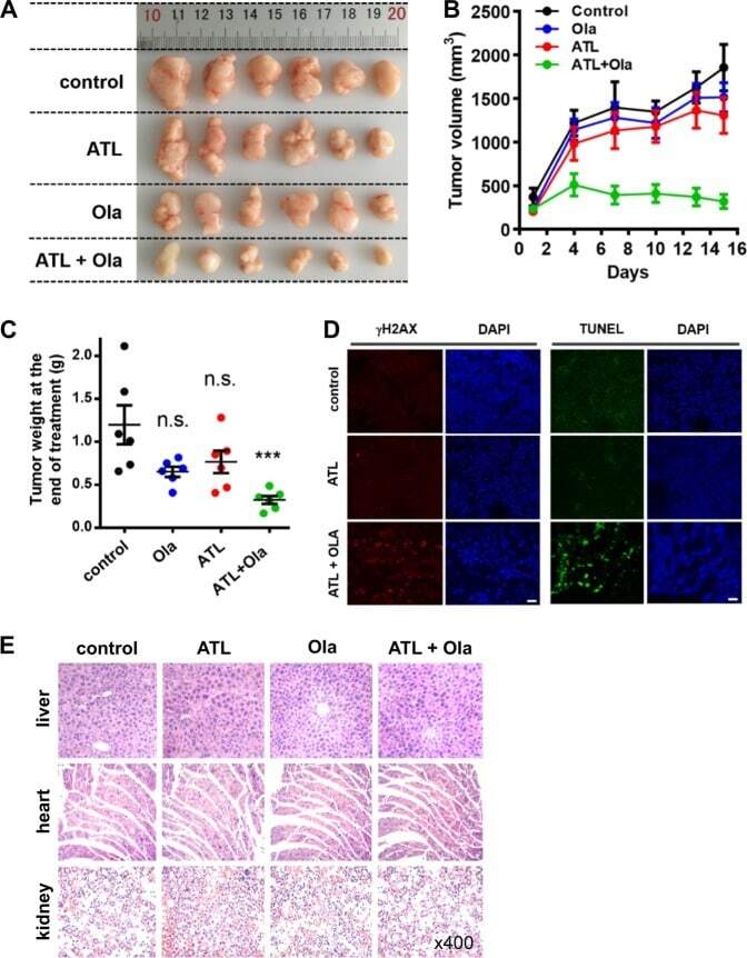
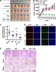
- Fig. 7 Coadministration of ATL and olaparib induces regression of tumor xenografts. PC-3 cells (2 x 10 6 ) in 1:1 matrigel were inoculated subcutaneously into the left flanks of male athymic BALB/c nude mice. When the tumor volume reached approximately 150 mm 3 (15 days after inoculation), mice were treated once daily with 50 mg/kg ATL oral gavage or 50 mg/kg olaparib intraperitoneal injection or both for 15 days. a Photograph of tumors dissected out from each mouse at the time of study termination. b Tumor volumes measured on the indicated days of treatment. Results were shown as mean +- SD. c Tumor weight measured at the end of the study. d Representative images of immunohistochemical staining of gammaH2AX and TUNEL in tumor tissues (scale bar: 20 mum). e Hematoxylin-eosin staining of liver, heart, and kidney tissue sections (magnification: x400). n.s. not significant, *** p < 0.001 vs. vehicle control.
- Conjugate
- Green dye