 Explore
 Validate
 Learn
Learn Western blot
 Immunoprecipitation
Antibody data
- Antibody Data
- Antigen structure
- References [3]
- Comments [0]
- Validations
- Western blot [11]
- Immunocytochemistry [2]
- Immunohistochemistry [6]
- Other assay [4]
Submit
Validation data
Reference
Comment
Report error
- Product number
- PA5-28778 - Provider product page

- Provider
- Invitrogen Antibodies
- Product name
- Histone H2A.X Polyclonal Antibody
- Antibody type
- Polyclonal
- Antigen
- Synthetic peptide
- Description
- Recommended positive controls: 293T, HepG2, HCT116, HCT116 (30uM cisplatin for 24hr), Neuro 2A, C8D30, NIH-3T3, Raw264.7, C2C12, mouse brain, rat brain, PC-12, Rat2, 4T1. Predicted reactivity: Rhesus Monkey (100%). Store product as a concentrated solution. Centrifuge briefly prior to opening the vial.
- Reactivity
- Human, Mouse, Rat
- Host
- Rabbit
- Isotype
- IgG
- Vial size
- 100 µL
- Concentration
- 1.41 mg/mL
- Storage
- Store at 4°C short term. For long term storage, store at -20°C, avoiding freeze/thaw cycles.
Submitted references The gut microbiota metabolite urolithin A inhibits NF-κB activation in LPS stimulated BMDMs.
Rapamycin-PLGA microparticles prevent senescence, sustain cartilage matrix production under stress and exhibit prolonged retention in mouse joints.
Chronic Intermittent Hypoxia Triggers a Senescence-like Phenotype in Human White Preadipocytes.
Abdelazeem KNM, Kalo MZ, Beer-Hammer S, Lang F
Scientific reports 2021 Mar 29;11(1):7117
Scientific reports 2021 Mar 29;11(1):7117
Rapamycin-PLGA microparticles prevent senescence, sustain cartilage matrix production under stress and exhibit prolonged retention in mouse joints.
Dhanabalan KM, Gupta VK, Agarwal R
Biomaterials science 2020 Aug 7;8(15):4308-4321
Biomaterials science 2020 Aug 7;8(15):4308-4321
Chronic Intermittent Hypoxia Triggers a Senescence-like Phenotype in Human White Preadipocytes.
Polonis K, Becari C, Chahal CAA, Zhang Y, Allen AM, Kellogg TA, Somers VK, Singh P
Scientific reports 2020 Apr 22;10(1):6846
Scientific reports 2020 Apr 22;10(1):6846
No comments: Submit comment
Supportive validation
- Submitted by
- Invitrogen Antibodies (provider)
- Main image

- Experimental details
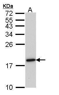
- Western blot analysis of Histone H2A.x using 30 µg of MOLT4 lysate. Samples were loaded onto a 15% SDS-PAGE gel and probed with a Histone H2A.x polyclonal antibody (Product # PA5-28778) at a dilution of 1:1000.
- Submitted by
- Invitrogen Antibodies (provider)
- Main image

- Experimental details
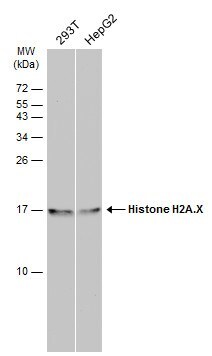
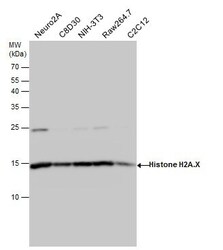
- Western blot analysis of Histone H2A.X in Various whole cell extracts (30 µg). Samples were separated by 15% SDS-PAGE and the membrane was probed with Histone H2A.X Polyclonal antibody (Product # PA5-28778) at a dilution of 1:500.

- Submitted by
- Invitrogen Antibodies (provider)
- Main image

- Experimental details
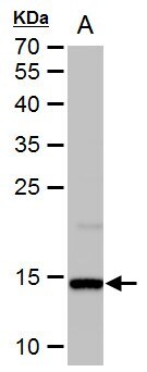
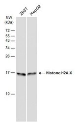
- Western Blot analysis of Histone H2A.X was performed by separating 30 µg of various whole cell extracts by 15% SDS-PAGE. Proteins were transferred to a membrane and probed with a Histone H2A.X Polyclonal Antibody (Product # PA5-28778) at a dilution of 1:1000 and a HRP-conjugated anti-rabbit IgG secondary antibody.
- Submitted by
- Invitrogen Antibodies (provider)
- Main image

- Experimental details
- Western Blot analysis of Histone H2A.X was performed by separating 30 µg of untreated (–) and treated (+) HCT116 whole cell extracts by 15% SDS-PAGE. Proteins were transferred to a membrane and probed with a Histone H2A.X Polyclonal Antibody (Product # PA5-28778) at a dilution of 1:10000.

- Submitted by
- Invitrogen Antibodies (provider)
- Main image

- Experimental details
- Western Blot analysis of Histone H2A.X was performed by separating 50 µg of mouse brain extracts by 12% SDS-PAGE. Proteins were transferred to a membrane and probed with a Histone H2A.X Polyclonal Antibody (Product # PA5-28778) at a dilution of 1:1000. The HRP-conjugated anti-rabbit IgG antibody was used to detect the primary antibody.
- Submitted by
- Invitrogen Antibodies (provider)
- Main image
- Experimental details
- Western Blot using Histone H2A. X Polyclonal Antibody (Product # PA5-28778). Various whole cell extracts (30 µg) were separated by 15% SDS-PAGE, and the membrane was blotted with Histone H2A. X Polyclonal Antibody (Product # PA5-28778) diluted at 1:1,000. The HRP-conjugated anti-rabbit IgG antibody was used to detect the primary antibody.

- Submitted by
- Invitrogen Antibodies (provider)
- Main image

- Experimental details
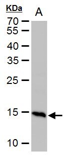
- Histone H2A. X Polyclonal Antibody detects Histone H2A. X protein by western blot analysis. A. 50 µg rat brain extract.12% SDS-PAGE. Histone H2A. X Polyclonal Antibody (Product # PA5-28778) dilution: 1:1,000. The HRP-conjugated anti-rabbit IgG antibody was used to detect the primary antibody.
- Submitted by
- Invitrogen Antibodies (provider)
- Main image

- Experimental details
- Western Blot using Histone H2A. X Polyclonal Antibody (Product # PA5-28778). Various whole cell extracts (30 µg) were separated by 15% SDS-PAGE, and the membrane was blotted with Histone H2A. X Polyclonal Antibody (Product # PA5-28778) diluted at 1:500. The HRP-conjugated anti-rabbit IgG antibody was used to detect the primary antibody.

- Submitted by
- Invitrogen Antibodies (provider)
- Main image

- Experimental details
- Western blot analysis was performed on acid extracts (20 µg lysate) of HeLa (Lane 1), K-562 (Lane 2), MDA-MB-231 (Lane 3), COS-7 (Lane 4) and A549 (Lane 5). The blot was probed with Anti-Histone H2A.X Polyclonal Antibody (Product # PA5-28778, 1:1000 dilution) and detected by chemiluminescence using Goat anti-Rabbit IgG (H+L) Superclonal™ Secondary Antibody, HRP conjugate (Product # A27036, 0.25 µg/mL, 1:4000 dilution). A 17 kDa band corresponding to H2A.X was observed across the cell lines tested.

Supportive validation
- Submitted by
- Invitrogen Antibodies (provider)
- Main image
- Experimental details
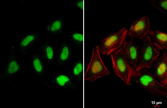
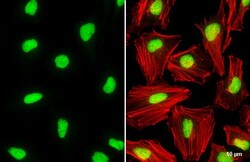
- Immunocytochemistry-Immunofluorescence analysis of Histone H2A.X was performed in HeLa cells fixed in 4% paraformaldehyde at RT for 15 min. Green: Histone H2A.X Polyclonal Antibody (Product # PA5 28778) diluted at 1:500. Red: phalloidin, a cytoskeleton marker. Scale bar = 10 µm.
- Submitted by
- Invitrogen Antibodies (provider)
- Main image
- Experimental details
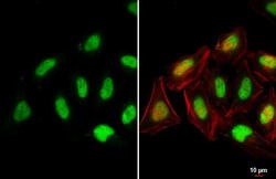
- Histone H2A. X Polyclonal Antibody detects Histone H2A. X protein at nucleus by immunofluorescent analysis. Sample: HeLa cells were fixed in 4% paraformaldehyde at RT for 15 min. Green: Histone H2A. X stained by Histone H2A. X Polyclonal Antibody (Product # PA5-28778) diluted at 1:2,000. Red: phalloidin, a cytoskeleton marker, diluted at 1:200. Scale bar= 10 µm.

Supportive validation
- Submitted by
- Invitrogen Antibodies (provider)
- Main image

- Experimental details
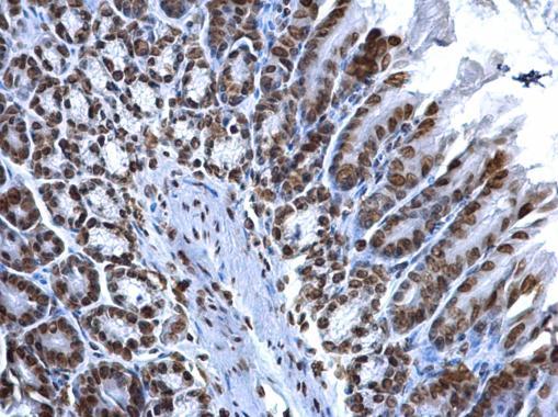
- Immunohistochemistry (Paraffin) analysis of Histone H2A.X was performed in paraffin-embedded human esophagus cancer tissue using Histone H2A.X Polyclonal Antibody (Product # PA5-28778) at a dilution of 1:500.

- Submitted by
- Invitrogen Antibodies (provider)
- Main image

- Experimental details
- Immunohistochemistry (Paraffin) analysis of Histone H2A.X was performed in paraffin-embedded human cervical cancer tissue using Histone H2A.X Polyclonal Antibody (Product # PA5-28778) at a dilution of 1:500.

- Submitted by
- Invitrogen Antibodies (provider)
- Main image

- Experimental details
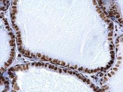
- Histone H2A. X Polyclonal Antibody detects Histone H2A. X protein at nucleus by immunohistochemical analysis. Sample: Paraffin-embedded human breast carcinoma. Histone H2A. X stained by Histone H2A. X Polyclonal Antibody (Product # PA5-28778) diluted at 1:500. Antigen Retrieval: Citrate buffer, pH 6.0, 15 min.

- Submitted by
- Invitrogen Antibodies (provider)
- Main image

- Experimental details
- Histone H2A. X Polyclonal Antibody detects Histone H2A. X protein at nucleus on mouse prostate by immunohistochemical analysis. Sample: Paraffin-embedded mouse prostate. Histone H2A. X Polyclonal Antibody (Product # PA5-28778) dilution: 1:500. Antigen Retrieval: EDTA based buffer, pH 8.0, 15 min.
- Submitted by
- Invitrogen Antibodies (provider)
- Main image
- Experimental details
- Histone H2A. X Polyclonal Antibody detects Histone H2A. X protein at nucleus on mouse prostate by immunohistochemical analysis. Sample: Paraffin-embedded mouse prostate. Histone H2A. X Polyclonal Antibody (Product # PA5-28778) dilution: 1:500. Antigen Retrieval: EDTA based buffer, pH 8.0, 15 min.

- Submitted by
- Invitrogen Antibodies (provider)
- Main image
- Experimental details
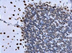
- Histone H2A. X Polyclonal Antibody detects Histone H2A. X protein at nucleus on rat hind brain by immunohistochemical analysis. Sample: Paraffin-embedded rat hind brain. Histone H2A. X Polyclonal Antibody (Product # PA5-28778) dilution: 1:500. Antigen Retrieval: EDTA based buffer, pH 8.0, 15 min.

Supportive validation
- Submitted by
- Invitrogen Antibodies (provider)
- Main image
- Experimental details
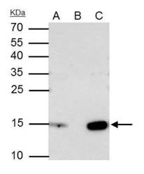
- Histone H2A. X Polyclonal Antibody immunoprecipitates H2AFX protein in IP experiments. IP samples: Jurkat whole cell extract. A. 40 µg Jurkat whole cell extract. B. Control with 4 µg of preimmune Rabbit IgG. C. Immunoprecipitation of H2AFX protein by 4 µg Histone H2A. X Polyclonal Antibody (Product # PA5-28778). 15 % SDS-PAGE. The immunoprecipitated H2AFX protein was detected by Histone H2A. X Polyclonal Antibody (Product # PA5-28778) diluted at 1:2,000.

- Submitted by
- Invitrogen Antibodies (provider)
- Main image
- Experimental details
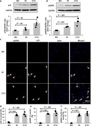
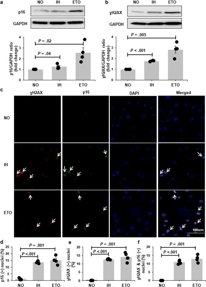
- Figure 2 P16 and gammaH2AX expression is increased with chronic exposure to intermittent hypoxia (IH). Representative Western bolts and graphs showing upregulation of p16 ( a ) and gammaH2AX ( b ) protein expression with IH treatment. Representative confocal images showing increased nuclear localization of p16 (green) and gammaH2AX (red) in cells exposed to IH ( c ). Nuclei are counterstained blue (DAPI). White arrows indicate positive nuclei. Quantitation of cells positive for nuclear p16 ( d ), gammaH2AX ( e ) and p16&gammaH2AX ( f ) in preadipocytes grown in continuous normoxia (NO) versus cells grown with intermittent exposure to hypoxia (n = 4 independent experiments). Cells treated with etopside (ETO) were used as positive control. Data are presented as mean +- SEM. P -values determined by one-tailed paired t-test compared to the NO control.
- Submitted by
- Invitrogen Antibodies (provider)
- Main image
- Experimental details
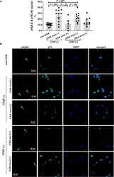
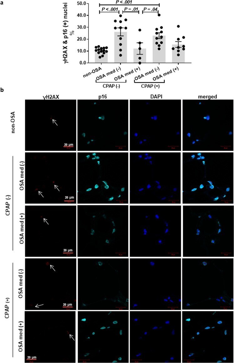
- Figure 8 Adipose tissue of OSA patients show increased expression of nuclear p16 and gammaH2AX. OSA was associated with a higher prevalence of cells with senescence-like phenotype in subcutaneous adipose tissue as determined by nuclear localization of both gammaH2AX and p16 (a) . The prevalence of dual positive nuclei was not attenuated by CPAP usage but was altered by concomitant use of medications. Representative images of immunofluorescence staining with white arrows indicating gammaH2AX positive nuclei (b) . OSA - obstructive sleep apnea, CPAP - continuous positive airway pressure, med - statins, aspirin and/or renin-angiotensin system inhibitors, (+/-) - present or absent. Data are presented as mean +- SEM. P -values determined by linear regression model adjusted for age and BMI.
- Submitted by
- Invitrogen Antibodies (provider)
- Main image
- Experimental details
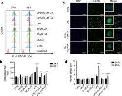
- Figure 3 Urolithin A minimized the intracellular calcium concentration and ameliorated DSBs in LPS-stimulated murine BMDMs. ( a , b ) Murine BMDMs were stimulated with 1 ug/ml LPS in the presence or absence of UA (25 uM or 50 uM) for 24 h or 48 h. ( a ) Representative FACS histograms showing the effect of UA on stimulated BMDMs after 24 h and 48 h. The reference line was set at the peak of the control. ( b ) Arithmetic means +- SEM (n = 5-7) show a significant difference in intracellular Ca 2+ concentration between control and treated groups after 24 h and 48 h. The unstimulated BMDMs were used as untreated control. DMSO was used as negative control. The intracellular Ca 2+ was measured with flow cytometry. (c) Murine BMDMs were stimulated by 1 ug/ml of LPS with or without UA (25 uM or 50 uM) for 2 h (images not shown) and 48 h. After 48 h, LPS induced DSBs indicated by a prominent green immunofluorescence for Ser139-phosphorylated H2AX. Treatment with UA recorded a remarkable decrease in number of gammaH2AX foci. Nuclei were counterstained with DAPI (blue). The magnifications are 20-fold and scale bar represents 50 mum. The unstimulated and untreated BMDMs were used as control. DMSO was used as negative control. ( d ) Graph indicates the number of gammaH2AX foci per cell after indicated time points. Representative images and arithmetic means +- SEM from four independent experiments (600 cells were counted). Two way ANOVA was used and ***( p < 0.001) indicate statistically si
