 Explore
 Validate
 Learn
Learn Western blot
Antibody data
- Antibody Data
- Antigen structure
- References [0]
- Comments [0]
- Validations
- Western blot [2]
- Immunocytochemistry [1]
- Immunohistochemistry [4]
- Other assay [1]
Submit
Validation data
Reference
Comment
Report error
- Product number
- PA5-58711 - Provider product page

- Provider
- Invitrogen Antibodies
- Product name
- alpha Tubulin Polyclonal Antibody
- Antibody type
- Polyclonal
- Antigen
- Recombinant full-length protein
- Description
- Immunogen sequence: LEHGIQPDGQ MPSDKTIGGG DDSFNTFF Highest antigen sequence identity to the following orthologs: Mouse - 100%, Rat - 100%.
- Reactivity
- Human, Mouse, Rat
- Host
- Rabbit
- Isotype
- IgG
- Vial size
- 100 µL
- Concentration
- 0.15 mg/mL
- Storage
- Store at 4°C short term. For long term storage, store at -20°C, avoiding freeze/thaw cycles.
No comments: Submit comment
Supportive validation
- Submitted by
- Invitrogen Antibodies (provider)
- Main image

- Experimental details
- Western blot analysis of alpha Tubulin in Lane 1: NIH-3T3 cell lysate (Mouse embryonic fibroblast cells); Lane 2: NBT-II cell lysate (Rat Wistar bladder tumour cells). Samples were probed using an Alpha Tubulin Polyclonal Antibody (Product # PA5-58711).

- Submitted by
- Invitrogen Antibodies (provider)
- Main image

- Experimental details
- Western blot analysis of alpha Tubulin in human cell line RT-4 and human cell line U-251 MG using a alpha Tubulin Polyclonal Antibody (Product # PA5-58711).

Supportive validation
- Submitted by
- Invitrogen Antibodies (provider)
- Main image
- Experimental details
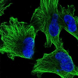
- Immunofluorescent staining of alpha Tubulin in human cell line U-251 MG using an Alpha Tubulin Polyclonal Antibody (Product # PA5-58711) shows localization to microtubules.

Supportive validation
- Submitted by
- Invitrogen Antibodies (provider)
- Main image

- Experimental details
- Immunohistochemical staining of alpha Tubulin in human cerebral cortex using alpha Tubulin Polyclonal Antibody (Product # PA5-58711) shows strong cytoplasmic positivity in neuropil.

- Submitted by
- Invitrogen Antibodies (provider)
- Main image

- Experimental details
- Immunohistochemical staining of alpha Tubulin in human testis using alpha Tubulin Polyclonal Antibody (Product # PA5-58711) shows strong cytoplasmic positivity in cells in seminiferous ducts.

- Submitted by
- Invitrogen Antibodies (provider)
- Main image

- Experimental details
- Immunohistochemical staining of alpha Tubulin in human fallopian tube using alpha Tubulin Polyclonal Antibody (Product # PA5-58711) shows strong cytoplasmic positivity in glandular cells.

- Submitted by
- Invitrogen Antibodies (provider)
- Main image

- Experimental details
- Immunohistochemical staining of alpha Tubulin in human liver using alpha Tubulin Polyclonal Antibody (Product # PA5-58711) shows no positivity in hepatocytes as expected.

Supportive validation
- Submitted by
- Invitrogen Antibodies (provider)
- Main image

- Experimental details
- Figure EV1 The addition of antibodies reduces viability and enhances TKI-induced apoptosis PC9 cells (5 x 10 3 ) were seeded in 96-well plates and later treated for 72 h with two different concentrations of EGFR-specific TKIs (erlotinib or osimertinib, at 10 or 100 nM; afatinib, at 1 or 10 nM), either alone or combined with 2XmAbs (cetuximab and trastuzumab, each at 5 mug/ml). Cell viability was assessed using the MTT (3-(4,5-dimethylthiazol-2-yl)-2,5-diphenyltetrazolium bromide) assay. Results are presented as mean + SEM of two independent experiments. PC9 cells were treated for 48 h with increasing concentrations of EGFR-specific TKIs (erlotinib or osimertinib at 10, 20, 40, 80, and 160 nM; afatinib at 1, 5, 10, 20 and 40 nM), either alone or in combination with 2XmAbs (cetuximab plus trastuzumab, each at 5 mug/ml). Protein extracts were resolved, blotted, and probed with antibodies specific to the indicated apoptosis markers. Tubulin (or GAPDH) was used as loading control. Signals (relative to control) were quantified and normalized to the signals of GAPDH or tubulin (numbers shown below each lane). PC9 cells were treated for 48 h with erlotinib (40 nM), osimertinib (40 nM), afatinib (10 nM), 2XmAbs (cetuximab and trastuzumab, each at 5 mug/ml), or combinations of mAbs and TKIs. Apoptosis was assayed using cytometry and the Annexin V/7-AAD kit (from BioLegend, Inc). The histogram shows the means + SEM of two experiments. Source data are available online for this figure.
