Explore
 Validate
 Learn
Learn Western blot
 ELISA
ELISAAntibody data
- Antibody Data
- Antigen structure
- References [0]
- Comments [0]
- Validations
- Western blot [1]
- Immunohistochemistry [1]
- Other assay [1]
Submit
Validation data
Reference
Comment
Report error
- Product number
- PA1-26809 - Provider product page

- Provider
- Invitrogen Antibodies
- Product name
- Fibrinogen Polyclonal Antibody
- Antibody type
- Polyclonal
- Antigen
- Other
- Reactivity
- Human
- Host
- Goat
- Isotype
- IgG
- Vial size
- 100 µL
- Concentration
- 90 mg/mL
- Storage
- Store at 4°C short term. For long term storage, store at -20°C, avoiding freeze/thaw cycles.
No comments: Submit comment
Supportive validation
- Submitted by
- Invitrogen Antibodies (provider)
- Main image
- Experimental details
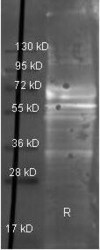
- Western Blot of Goat anti-Fibrinogen Polyclonal Antibody (Product # PA1-26809). Lane 1: Fibrinogen under reducing conditions. Load: 1 µg per lane. Primary antibody: Fibrinogen Polyclonal Antibody at 1:3,000 for overnight at 4ºC.

Supportive validation
- Submitted by
- Invitrogen Antibodies (provider)
- Main image
- Experimental details
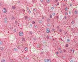
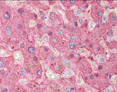
- Immunohistochemistry (Paraffin) of Fibrinogen was performed in formalin fixed, paraffin embedded human liver tissue. The Fibrinogen Polyclonal Antibody (Product # PA1-26809) was used as a primary antibody at a dilution of 1:500 for 1 hour at room temperature, and a peroxidase goat antibody was used as a secondary at a dilution of 1:10,000 for 45 min at RT. Localization: Fibrinogen is localized in plasma. Staining: Fibrinogen as precipitated red signal with hematoxylin purple nuclear counterstain.
Supportive validation
- Submitted by
- Invitrogen Antibodies (provider)
- Main image
- Experimental details
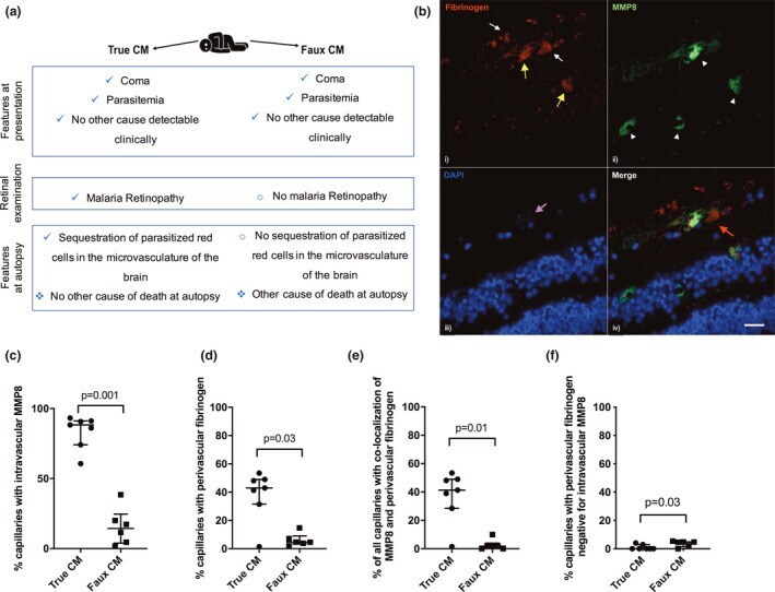
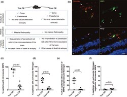
- Figure 1 Matrix metallopeptidase 8 (MMP8) co-localises with fibrinogen leak in the retinal capillaries in 'true cerebral malaria'. (a) Illustration of the similarities and differences between 'true'- and 'faux'-CM subjects. (b) Representative image showing co-localisation of intravascular MMP8 and perivascular fibrinogen staining in the retina of a subject with true cerebral malaria (CM). (i) Fibrinogen (red) staining was seen intravascularly (yellow arrows), likely in areas of microthrombus formation, and immediately outside of capillaries (white arrows), indicating vascular leak. (ii) MMP8 (green) staining was observed intravascularly (arrowheads). (iii) DAPI (blue) stains nuclei, magenta arrow points to a capillary with heavy sequestration of parasitised erythrocytes (parasite nuclei visible as fine blue dots). (iv) Merged image shows co-localisation of intravascular MMP8 and fibrinogen leak (red arrow) around one of the capillaries. Scale bar: 25 um. (c-f) Quantification of intravascular MMP8 and perivascular fibrinogen staining in the retina of subjects with 'true'- and 'faux'-CM. (c) Percentage of observed capillaries with intravascular MMP8 staining. (d) Percentage of observed capillaries with perivascular fibrinogen staining. (e) Percentage of all capillaries in each sample, with co-localisation of intravascular MMP8 and perivascular fibrinogen. (f) Percentage of capillaries with perivascular fibrinogen staining which are negative for intravascular MMP8. Bars show med