Explore
 Validate
 Learn
Learn Western blot
 ELISA
ELISAAntibody data
- Antibody Data
- Antigen structure
- References [1]
- Comments [0]
- Validations
- Western blot [1]
- Immunocytochemistry [4]
- Immunohistochemistry [3]
- Other assay [2]
Submit
Validation data
Reference
Comment
Report error
- Product number
- MA5-15507 - Provider product page

- Provider
- Invitrogen Antibodies
- Product name
- Cytokeratin Pan Monoclonal Antibody (7H8)
- Antibody type
- Monoclonal
- Antigen
- Purifed from natural sources
- Description
- MA5-15507 targets Cytokeratin(Pan) in IF and IHC applications and shows reactivity with Human samples. The MA5-15507 immunogen is purified recombinant fragment of Cytokeratin 5 expressed in E. Coli.
- Reactivity
- Human
- Host
- Mouse
- Isotype
- IgG
- Antibody clone number
- 7H8
- Vial size
- 100 µL
- Concentration
- Conc. Not Determined
- Storage
- Store at 4°C short term. For long term storage, store at -20°C, avoiding freeze/thaw cycles.
Submitted references Neurodegeneration-Associated Proteins in Human Olfactory Neurons Collected by Nasal Brushing.
Brozzetti L, Sacchetto L, Cecchini MP, Avesani A, Perra D, Bongianni M, Portioli C, Scupoli M, Ghetti B, Monaco S, Buffelli M, Zanusso G
Frontiers in neuroscience 2020;14:145
Frontiers in neuroscience 2020;14:145
No comments: Submit comment
Supportive validation
- Submitted by
- Invitrogen Antibodies (provider)
- Main image
- Experimental details
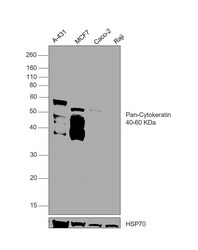
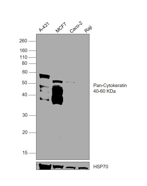
- Western blot was performed using Anti-Cytokeratin Pan Monoclonal Antibody (7H8) (Product # MA5-15507) and a 40-60 kDa band corresponding to Cytokeratin Pan was observed across in all tested cell lines, except Raji. Whole cell extracts (30 µg lysate) of A-431 (Lane 1), MCF7 (Lane 2), Caco-2 (Lane 3), Raji (Lane 4) were electrophoresed using NuPAGE™ 10% Bis-Tris Protein Gel (Product # NP0301BOX). Resolved proteins were then transferred onto a nitrocellulose membrane (Product # IB23002) by iBlot® 2 Dry Blotting System (Product # IB21001). The blot was probed with the primary antibody (1:1000 dilution) and detected by chemiluminescence with Goat anti-Mouse IgG (H+L) Superclonal™ Recombinant Secondary Antibody, HRP (Product # A28177,1:10000) using the iBright™ FL1500 Imaging System (Product # A44115). Chemiluminescent detection was performed using SuperSignal™ West Pico PLUS Chemiluminescent Substrate (Product # 34580).
Supportive validation
- Submitted by
- Invitrogen Antibodies (provider)
- Main image
- Experimental details
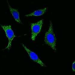
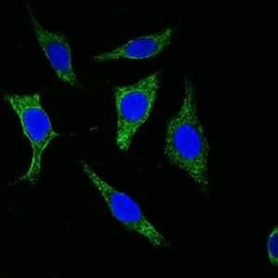
- Immunofluorescence analysis of methanol-fixed Eca-109 cells using Cytokeratin (Pan) monoclonal antibody (Product # MA5-15507) (Green). Blue: DRAQ5 fluorescent DNA dye.

- Submitted by
- Invitrogen Antibodies (provider)
- Main image
- Experimental details
- Immunofluorescence analysis of methanol-fixed Eca-109 cells using Cytokeratin (Pan) monoclonal antibody (Product # MA5-15507) (Green). Blue: DRAQ5 fluorescent DNA dye.

- Submitted by
- Invitrogen Antibodies (provider)
- Main image

- Experimental details
- Immunofluorescence analysis of methanol-fixed Eca-109 cells using Cytokeratin (Pan) monoclonal antibody (Product # MA5-15507) (Green). Blue: DRAQ5 fluorescent DNA dye.

- Submitted by
- Invitrogen Antibodies (provider)
- Main image
- Experimental details
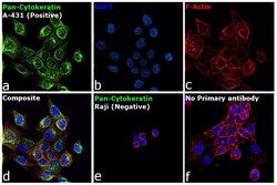
- Immunofluorescence analysis of Cytokeratin Pan was performed using 70% confluent log phase A-431 cells. The cells were fixed and permeabilized with ice-cold acetone at 4°C for 5 minutes and blocked with 2% BSA for 1 hour at room temperature. The cells were labeled with Cytokeratin Pan Monoclonal Antibody (7H8) (Product # MA5-15507, 1:100 dilution) in 0.1% BSA, incubated at 4 degree celsius overnight and then labeled with Donkey anti-Mouse IgG (H+L) Highly Cross-Adsorbed Secondary Antibody, Alexa Fluor Plus 488 (Product # A32766, 1:2000 dilution), for 45 minutes at room temperature (Panel a: Green). Nuclei (Panel b: Blue) were stained with ProLong™ Diamond Antifade Mountant with DAPI (Product # P36962). F-actin (Panel c: Red) was stained with Rhodamine Phalloidin (Product # R415, 1:300 dilution). Panel d represents the merged image showing cytoskeletal localization. Panel e represents Raji cells showing no expression of cytokeratin. Panel f represents control cells with no primary antibody to assess background. The images were captured at 60X magnification.

Supportive validation
- Submitted by
- Invitrogen Antibodies (provider)
- Main image
- Experimental details
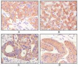
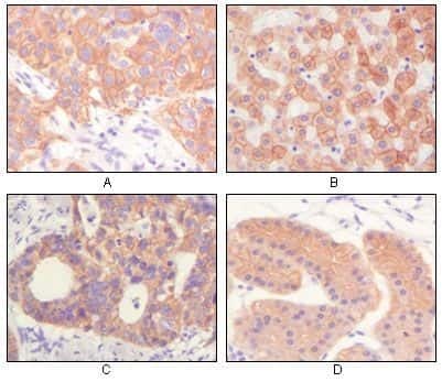
- Immunohistochemical analysis of paraffin-embedded human lung squamous cell carcinoma (A), normal hepatocyte (B), colon adenocacinoma, normal stomach tissue (D), showing cytoplasmic and membrane localization using Cytokeratin (Pan) monoclonal antibody (Product # MA5-15507) followed with DAB staining.
- Submitted by
- Invitrogen Antibodies (provider)
- Main image

- Experimental details
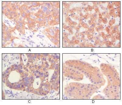
- Immunohistochemical analysis of paraffin-embedded human lung squamous cell carcinoma (A), normal hepatocyte (B), colon adenocacinoma, normal stomach tissue (D), showing cytoplasmic and membrane localization using Cytokeratin (Pan) monoclonal antibody (Product # MA5-15507) followed with DAB staining.
- Submitted by
- Invitrogen Antibodies (provider)
- Main image
- Experimental details
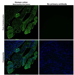
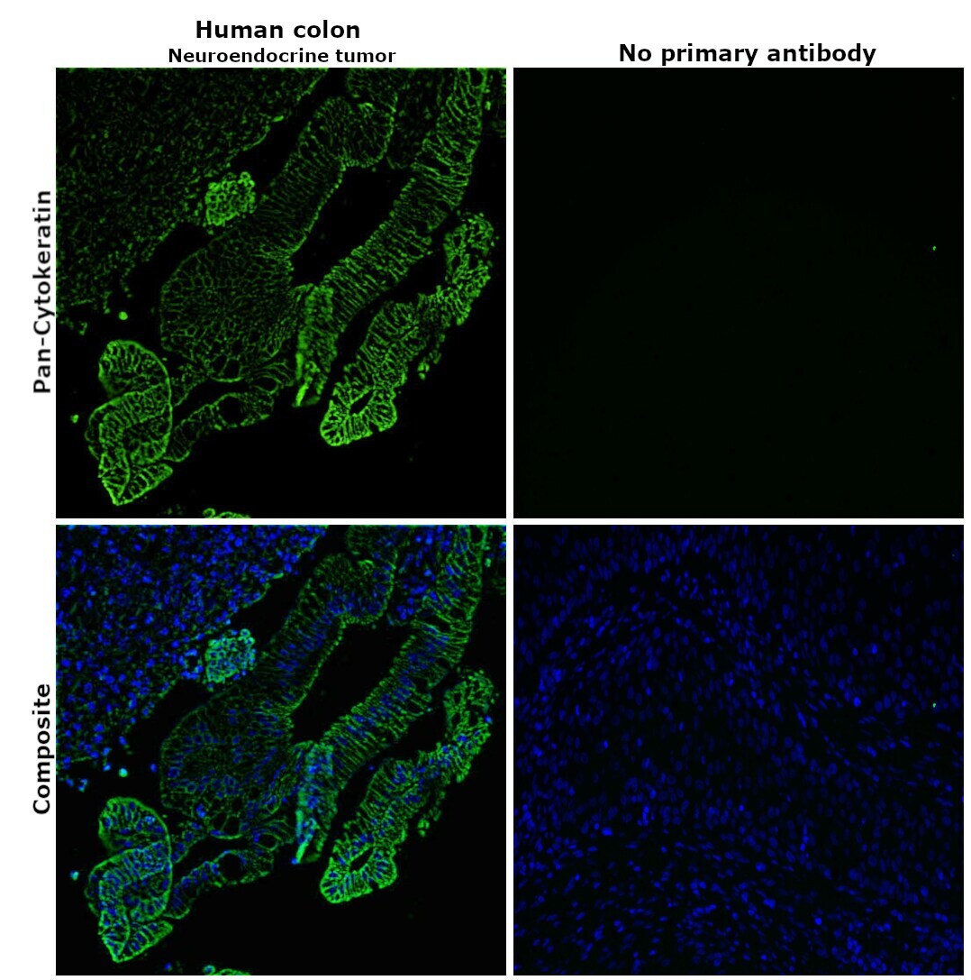
- Immunohistochemical analysis of pan Cytokeratin was performed using formalin-fixed paraffin-embedded human colon (neuroendocrine tumor) tissue sections. To expose the target protein, heat-induced epitope retrieval was performed on de-paraffinized sections using eBioscience™ IHC Antigen Retrieval Solution - Low pH (10X) (Product # 00-4955-58) diluted to 1X solution in water in a decloaking chamber at 110 degree Celsius for 15 minutes. Following antigen retrieval, the sections were blocked with 2% normal goat serum in 1X PBS for 45 minutes at room temperature and then probed with or without Cytokeratin Pan Monoclonal Antibody (7H8) (Product # MA5-15507) at 1:100 dilution in 0.1% normal goat serum overnight at 4 degree Celsius in a humidified chamber. Detection was performed using Goat anti-Mouse IgG (H+L) Highly Cross-Adsorbed Secondary Antibody, Alexa Fluor Plus 488 (Product # A32723) at a dilution of 1:2000 in 0.1% normal goat serum for 45 minutes at room temperature. Nuclei were stained with DAPI (Product # D1306) and the sections were mounted using ProLong™ Glass Antifade Mountant (Product # P36984). The images were captured at 20X magnification.
Supportive validation
- Submitted by
- Invitrogen Antibodies (provider)
- Main image
- Experimental details
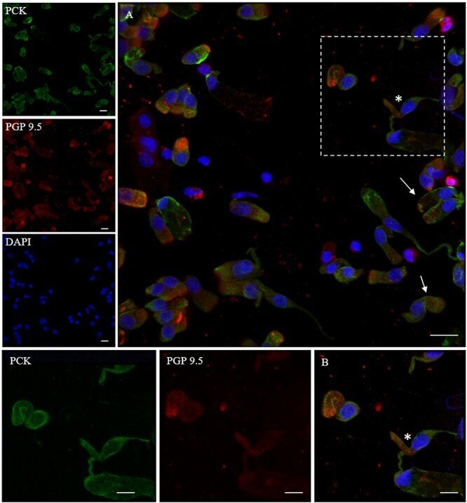
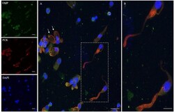
- FIGURE 2 Immunocytochemical analysis of a cytocentrifuged sample of olfactory mucosa (OM) using OMP (green) and PCK (red). While OMP stains round and non-neuronal-shaped cells, PCK preferentially stains the whole apical dendritic projection of olfactory neurons [outlined square ( A ) up to the cilia boundary (detail B )]. Interestingly, in ONs, the immunopositivity with PCK is distributed on the opposite side of that obtained with beta-tubulin III. In the other cells, PCK expression is distributed on the boundary of the cell body, all along the plasma membrane (arrows) of cells that have a round shape. Scale bar (A) : 20 mum. Scale bar (B) : 10 mum.

- Submitted by
- Invitrogen Antibodies (provider)
- Main image
- Experimental details
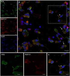
- FIGURE 3 Distribution pattern of PGP 9.5 and PCK. PGP 9.5 (red) shows a dotted positivity mostly in the cytosol of cells with sustentacular-like morphology and less intensely in olfactory neurons (*, square detail). PCK (green) positivity is also identified in the cytosolic compartment of olfactory neurons and on the plasma membrane of other cells with non-neuronal morphology (arrows). Scale bar (A) : 20 mum. Scale bar (B) : 10 mum.