Explore
 Validate
 Learn
Learn Western blot
 Immunoprecipitation
Antibody data
- Antibody Data
- Antigen structure
- References [1]
- Comments [0]
- Validations
- Western blot [3]
- Immunocytochemistry [1]
- Immunohistochemistry [1]
Submit
Validation data
Reference
Comment
Report error
- Product number
- PA5-17320 - Provider product page

- Provider
- Invitrogen Antibodies
- Product name
- gamma Catenin Polyclonal Antibody
- Antibody type
- Polyclonal
- Antigen
- Synthetic peptide
- Description
- It is not recommended to aliquot this antibody. This antibody is not cross-reactive with -beta-catenin.
- Reactivity
- Human, Mouse, Rat, Hamster
- Host
- Rabbit
- Isotype
- IgG
- Vial size
- 100 µL
- Concentration
- 32 µg/mL
- Storage
- -20°C
Submitted references Cardiomyocyte Expression of ZO-1 Is Essential for Normal Atrioventricular Conduction but Does Not Alter Ventricular Function.
Zhang J, Vincent KP, Peter AK, Klos M, Cheng H, Huang SM, Towne JK, Ferng D, Gu Y, Dalton ND, Chan Y, Li R, Peterson KL, Chen J, McCulloch AD, Knowlton KU, Ross RS
Circulation research 2020 Jul 3;127(2):284-297
Circulation research 2020 Jul 3;127(2):284-297
No comments: Submit comment
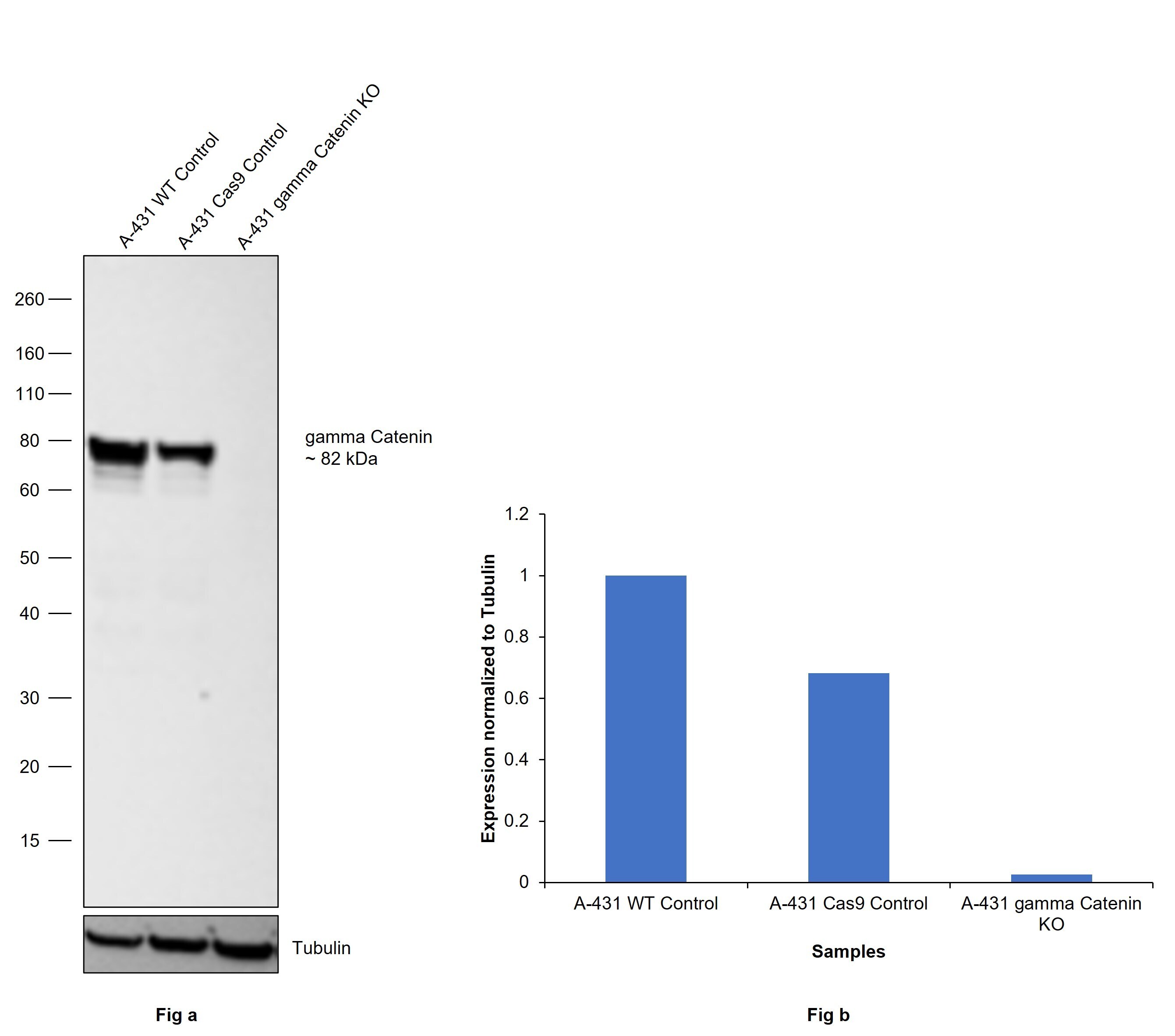
Supportive validation
- Submitted by
- Invitrogen Antibodies (provider)
- Main image

- Experimental details
- Knockout of gamma Catenin was achieved by CRISPR-Cas9 genome editing using LentiArray™ Lentiviral sgRNA (Product # A32042, Assay ID CRISPR680765_LV) and LentiArray Cas9 Lentivirus (Product # A32064). Western blot analysis of gamma Catenin was performed by loading 30 µg of A-431 Wild type (Lane 1), A-431 Cas9 (Lane 2) andA-431 gamma Catenin KO (Lane 3) whole cell extracts. The samples were electrophoresed using NuPAGE™ Novex™ 4-12% Bis-Tris Protein Gel (Product # NP0321BOX). Resolved proteins were then transferred onto a nitrocellulose membrane (Product # IB23001) by iBlot® 2 Dry Blotting System (Product # IB21001). The blot was probed with gamma Catenin Polyclonal Antibody (Product # PA5-17320, 1:1000 dilution) and Goat anti-Rabbit IgG (H+L) Superclonal™ Recombinant Secondary Antibody, HRP (Product # A27036, 1:10.000 dilution) using the iBright™ FL 1500 (Product # A44115). Chemiluminescent detection was performed using SuperSignal™ West Dura Extended Duration Substrate (Product # 34076). Loss of signal upon CRISPR mediated knockout (KO) using the LentiArray™ CRISPR product line confirms that antibody is specific to gamma Catenin.
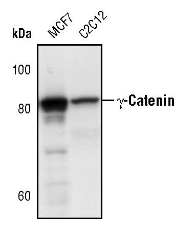
- Submitted by
- Invitrogen Antibodies (provider)
- Main image

- Experimental details
- Western blot analysis of gamma-Catenin in extracts from MCF-7 and C2C12 cells using gamma-Catenin polyclonal antibody (Product # PA5-17320).
- Submitted by
- Invitrogen Antibodies (provider)
- Main image
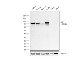
- Experimental details
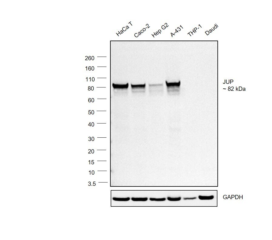
- Western blot was performed using Anti-gamma Catenin Polyclonal Antibody(Product # PA5-17320) and a 82 kDa band corresponding to gamma Catenin was observed across the cell lines tested except THP-1 and Daudi. Whole cell extracts (30 µg lysate) of HaCaT (Lane 1), Caco-2 (Lane 2), Hep G2 (Lane 3), A-431 (Lane 4), THP-1 (Lane 5) and Daudi (Lane 6) were electrophoresed using NuPAGE™ 4-12% Bis-Tris Protein Gel (Product # NP0321BOX). Resolved proteins were then transferred onto a Nitrocellulose membrane (Product # LC2001) by iBlot® 2 Dry Blotting System (Product # IB21001). The blot was probed with the primary antibody (1:1000 dilution) and detected by chemiluminescence with Goat anti-Rabbit IgG (H+L) Superclonal™ Recombinant Secondary Antibody, HRP (Product # A27036,1:4000 dilution) using the iBright FL 1000 (Product # A32752). Chemiluminescent detection was performed using Novex® ECL Chemiluminescent Substrate Reagent Kit (Product # WP20005).
Supportive validation
- Submitted by
- Invitrogen Antibodies (provider)
- Main image

- Experimental details
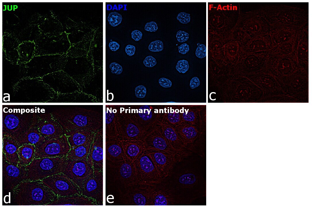
- Immunofluorescence analysis of gamma Catenin was performed using 70% confluent log phase HaCaT cells. The cells were fixed with 4% paraformaldehyde for 10 minutes, permeabilized with 0.1% Triton™ X-100 for 10 minutes, and blocked with 2% BSA for 45 minutes at room temperature. The cells were labeled with gamma Catenin Polyclonal Antibody (Product # PA5-17320) at 1:400 dilution in 0.1% BSA, incubated at 4 degree celsius overnight and then labeled with Goat anti-Rabbit IgG (H+L) Superclonal™ Recombinant Secondary Antibody, Alexa Fluor® 488 conjugate (Product # A27034), (1:2000 dilution), for 45 minutes at room temperature (Panel a: Green). Nuclei (Panel b:Blue) were stained with SlowFade® Gold Antifade Mountant with DAPI (Product # S36938). F-actin (Panel c: Red) was stained with Rhodamine Phalloidin (Product # R415, 1:300 dilution). Panel d represents the merged image showing membrane and cytoplasm localization. Panel e represents control cells with no primary antibody to assess background. The images were captured at 60X magnification.
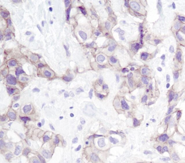
Supportive validation
- Submitted by
- Invitrogen Antibodies (provider)
- Main image

- Experimental details
- Immunohistochemical analysis of gamma-Catenin in paraffin-embedded human breastcarcinoma using a gamma-Catenin polyclonal antibody (Product # PA5-17320).