 Explore
 Validate
 Learn
Learn Western blot
Antibody data
- Antibody Data
- Antigen structure
- References [0]
- Comments [0]
- Validations
- Western blot [2]
- Immunocytochemistry [3]
- Immunohistochemistry [5]
- Other assay [5]
Submit
Validation data
Reference
Comment
Report error
- Product number
- MA5-13203 - Provider product page

- Provider
- Invitrogen Antibodies
- Product name
- Cytokeratin Pan Antibody Cocktail
- Antibody type
- Monoclonal
- Antigen
- Other
- Description
- This antibody is a broad-spectrum pan-keratin monoclonal antibody cocktail which recognizes keratin 4, 5, 6, 7, 8, 10, 13, 14, 18 and 19. This product can differentiate between epithelial and non-epithelial tumors. For staining of formalin-fixed tissue,digest sections wtih trypsin at 1 mg/mL in PBS, 10 min at 37ºC, or Protease XXV at 1 mg/mL in PBS for 5 min at 37ºC.
- Reactivity
- Human, Mouse
- Host
- Mouse
- Isotype
- IgG
- Antibody clone number
- PAN-CK
- Vial size
- 500 µL
- Concentration
- 0.2 mg/mL
- Storage
- 4° C
No comments: Submit comment
Supportive validation
- Submitted by
- Invitrogen Antibodies (provider)
- Main image

- Experimental details
- Western blot analysis of Cytokeratin Pan was performed by loading 25 µg of A431 (Lane 1), Hela (Lane 2), and PC12 cell lysates (Lane 3) and a molecular weight protein ladder onto an SDS polyacrylamide gel. Proteins were transferred to a PVDF membrane and blocked with a blocking buffer at 4ºC overnight. The membrane was probed with a Cytokeratin Pan monoclonal antibody (Product # MA5-13203) at a dilution of 1:2000 (A431 and Hela) and 1:1000 (PC12) overnight at 4°C, washed in TBST, and probed with an HRP-conjugated secondary antibody for 1 hr at room temperature in the dark. Chemiluminescent detection was performed using Pierce ECL Plus Western Blotting Substrate (Product # 32132). Results show a band at 40-67 kDa in A431 and Hela cell lines.

- Submitted by
- Invitrogen Antibodies (provider)
- Main image
- Experimental details
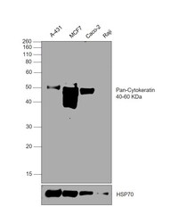
- Western blot was performed using Anti-Cytokeratin Pan Antibody Cocktail (Product # MA5-13203) and a 40-60 kDa band corresponding to Cytokeratin Pan was observed across in all tested cell lines, except Raji. Whole cell extracts (30 µg lysate) of A-431 (Lane 1), MCF7 (Lane 2), Caco-2 (Lane 3), Raji (Lane 4) were electrophoresed using NuPAGE™ 10% Bis-Tris Protein Gel (Product # NP0301BOX). Resolved proteins were then transferred onto a nitrocellulose membrane (Product # IB23002) by iBlot® 2 Dry Blotting System (Product # IB21001). The blot was probed with the primary antibody (1:1000 dilution) and detected by chemiluminescence with Goat anti-Mouse IgG (H+L) Superclonal™ Recombinant Secondary Antibody, HRP (Product # A28177,1:10000 dilution) using the iBright™ FL1500 Imaging System (Product # A44115). Chemiluminescent detection was performed using SuperSignal™ West Pico PLUS Chemiluminescent Substrate (Product # 34580).

Supportive validation
- Submitted by
- Invitrogen Antibodies (provider)
- Main image
- Experimental details
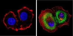
- Immunofluorescent analysis of Cytokeratin Pan (green) showing staining in the cytoplasm of MCF-7 cells. Formalin-fixed cells were permeabilized with 0.1% Triton X-100 in TBS for 5-10 minutes and blocked with 3% BSA-PBS for 30 minutes at room temperature. Cells were probed with a Cytokeratin Pan monoclonal antibody (Product # MA5-13203) in 3% BSA-PBS at a dilution of 1:100 and incubated overnight at 4 ºC in a humidified chamber. Cells were washed with PBST and incubated with a DyLight-conjugated secondary antibody in PBS at room temperature in the dark. F-actin (red) was stained with a fluorescent red phalloidin and nuclei (blue) were stained with Hoechst or DAPI. Images were taken at a magnification of 60x.

- Submitted by
- Invitrogen Antibodies (provider)
- Main image

- Experimental details
- Immunofluorescent analysis of Cytokeratin Pan (green) showing staining in the cytoplasm of HeLa cells. Formalin-fixed cells were permeabilized with 0.1% Triton X-100 in TBS for 5-10 minutes and blocked with 3% BSA-PBS for 30 minutes at room temperature. Cells were probed with a Cytokeratin Pan monoclonal antibody (Product # MA5-13203) in 3% BSA-PBS at a dilution of 1:100 and incubated overnight at 4 ºC in a humidified chamber. Cells were washed with PBST and incubated with a DyLight-conjugated secondary antibody in PBS at room temperature in the dark. F-actin (red) was stained with a fluorescent red phalloidin and nuclei (blue) were stained with Hoechst or DAPI. Images were taken at a magnification of 60x.

- Submitted by
- Invitrogen Antibodies (provider)
- Main image
- Experimental details
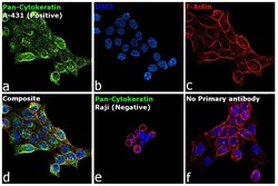
- Immunofluorescence analysis of Cytokeratin Pan was performed using 70% confluent log phase A-431 cells. The cells were fixed and permeabilized with ice-cold acetone at 4°C for 5 minutes, and blocked with 2% BSA for 1 hour at room temperature. The cells were labeled with Cytokeratin Pan Antibody Cocktail (Product # MA5-13203, 5 µg/mL) in 0.1% BSA, incubated at 4 degree celsius overnight and then labeled with Donkey anti-Mouse IgG (H+L) Highly Cross-Adsorbed Secondary Antibody, Alexa Fluor Plus 488 (Product # A32766, 1:2000 dilution), for 45 minutes at room temperature (Panel a: Green). Nuclei (Panel b: Blue) were stained with ProLong™ Diamond Antifade Mountant with DAPI (Product # P36962). F-actin (Panel c: Red) was stained with Rhodamine Phalloidin (Product # R415, 1:300 dilution). Panel d represents the merged image showing cytoskeletal localization. Panel e represents Raji cells showing no expression of cytokeratin. Panel f represents control cells with no primary antibody to assess background. The images were captured at 60X magnification.

Supportive validation
- Submitted by
- Invitrogen Antibodies (provider)
- Main image

- Experimental details
- Formalin-fixed, paraffin-embedded human skin stained with Keratin, Pan antibody using peroxidase-conjugate and AEC chromogen. Note cytoplasmic staining of epidermis.

- Submitted by
- Invitrogen Antibodies (provider)
- Main image

- Experimental details
- Immunohistochemistry analysis of Cytokeratin Pan showing positive staining in the cytoplasm of paraffin-treated Human colon carcinoma (right) compared with a negative control in the absence of primary antibody (left). To expose target proteins, antigen retrieval method was performed using 10mM sodium citrate (pH 6.0) microwaved for 8-15 min. Following antigen retrieval, tissues were blocked in 3% H2O2-methanol for 15 min at room temperature, washed with ddH2O and PBS, and then probed with a Cytokeratin Pan monoclonal antibody (Product # MA5-13203) diluted by 3% BSA-PBS at a dilution of 1:100 overnight at 4°C in a humidified chamber. Tissues were washed extensively PBST and detection was performed using an HRP-conjugated secondary antibody followed by colorimetric detection using a DAB kit. Tissues were counterstained with hematoxylin and dehydrated with ethanol and xylene to prep for mounting.

- Submitted by
- Invitrogen Antibodies (provider)
- Main image

- Experimental details
- Immunohistochemistry analysis of Cytokeratin Pan showing positive staining in the cytoplasm of paraffin-treated Human skin tissue (right) compared with a negative control in the absence of primary antibody (left). To expose target proteins, antigen retrieval method was performed using 10mM sodium citrate (pH 6.0) microwaved for 8-15 min. Following antigen retrieval, tissues were blocked in 3% H2O2-methanol for 15 min at room temperature, washed with ddH2O and PBS, and then probed with a Cytokeratin Pan monoclonal antibody (Product # MA5-13203) diluted by 3% BSA-PBS at a dilution of 1:200 overnight at 4°C in a humidified chamber. Tissues were washed extensively PBST and detection was performed using an HRP-conjugated secondary antibody followed by colorimetric detection using a DAB kit. Tissues were counterstained with hematoxylin and dehydrated with ethanol and xylene to prep for mounting.

- Submitted by
- Invitrogen Antibodies (provider)
- Main image

- Experimental details
- Immunohistochemistry analysis of Cytokeratin Pan showing positive staining in the cytoplasm of paraffin-treated Mouse skin tissue (right) compared with a negative control in the absence of primary antibody (left). To expose target proteins, antigen retrieval method was performed using 10mM sodium citrate (pH 6.0) microwaved for 8-15 min. Following antigen retrieval, tissues were blocked in 3% H2O2-methanol for 15 min at room temperature, washed with ddH2O and PBS, and then probed with a Cytokeratin Pan monoclonal antibody (Product # MA5-13203) diluted by 3% BSA-PBS at a dilution of 1:100 overnight at 4°C in a humidified chamber. Tissues were washed extensively PBST and detection was performed using an HRP-conjugated secondary antibody followed by colorimetric detection using a DAB kit. Tissues were counterstained with hematoxylin and dehydrated with ethanol and xylene to prep for mounting.

- Submitted by
- Invitrogen Antibodies (provider)
- Main image

- Experimental details
- Immunohistochemical analysis of pan Cytokeratin was performed using formalin-fixed paraffin-embedded human colon (neuro-endocrine tumor) tissue sections. To expose the target protein, heat-induced epitope retrieval (HIER) was performed on de-paraffinized sections using eBioscience™ IHC Antigen Retrieval Solution - Low pH (10X) (Product # 00-4955-58) diluted to 1X solution in water in a decloaking chamber at 110 degree Celsius for 20 minutes. Following antigen retrieval, the sections were blocked with 2% normal goat serum in 1X PBS for 45 minutes at room temperature and then probed with or without Cytokeratin Pan Antibody Cocktail (Product # MA5-13203) at 2 µg/mL in 0.1% normal goat serum overnight at 4 degree Celsius in a humidified chamber. Detection was performed using Goat anti-Mouse IgG (H+L) Highly Cross-Adsorbed Secondary Antibody, Alexa Fluor Plus 488 (Product # A32723) at a dilution of 1:2000 in 0.1% normal goat serum for 45 minutes at room temperature. Nuclei were stained with DAPI (Product # D1306) and the sections were mounted using ProLong™ Glass Antifade Mountant (Product # P36984). The images were captured on EVOS™ M7000 Imaging System (Product # AMF7000) at 20X magnification.

Supportive validation
- Submitted by
- Invitrogen Antibodies (provider)
- Main image

- Experimental details
- NULL

- Submitted by
- Invitrogen Antibodies (provider)
- Main image

- Experimental details
- Figure 3 Krtap5-5 stabilizes the keratin cytoskeleton. ( a ) Phalloidin staining for F-actin reveals cytoskeletal dynamics in Krtap5-5 knockdown lines. ( b ) Confocal imaging of vimentin intermediate filaments. ( c ) Confocal imaging of keratin intermediate filaments. Scale bars are 10 mum. ( d ) Western blot for pan-keratin in the control vs Krtap5-5 knockdown cell lines. ( e , f ) Keratin 14 ( e ) and Keratin 18 ( f ) mRNA expression in Krtap5-5 knockdown lines. *** p

- Submitted by
- Invitrogen Antibodies (provider)
- Main image

- Experimental details
- Figure 11 Expression of Vimentin, Pan-cytokeratin, beta-catenin and CD44 in cells disaggregated from T88 and T93 spheroids. Confocal images of cells disaggregated from T88 and T93 spheroids obtained in medium containing (LiCl) or not containing (untreated) LiCl. Cells were dual-stained with anti-Vimentin (red)/anti-Pan-cytokeratin (green) and anti-beta-catenin (red)/anti-CD44 (green) antibodies. Nuclei were counterstained with DAPI (blue).

- Submitted by
- Invitrogen Antibodies (provider)
- Main image

- Experimental details
- Fig 2 Histopathology analysis of WT, HET, and DKO tumors. (A) Representative sections of tissue form DKO mice, as well as both HET and WT at 25 and ~40 weeks. Disease stage was determined by pathologist review of H&E sections, with sarcomatoid being confirmed with a negative pan-cytokeratin stain. Bar = 100mum. Asterisks denote small patches of SMA-positivity. (B) Histopathology analysis from H&E and cyokeratin staining in (A), (*p>0.05; Chi-Square with Bonferroni correction).

- Submitted by
- Invitrogen Antibodies (provider)
- Main image

- Experimental details
- Figure 2. Morphologic characteristics and antigenic marker expression of the novel DDCS2 cell line. (A) DDCS2 cells display spindle to polygonal shape in vitro under light microscopy (200x, scale bar: 50 mum). (B) The growth curves of DDCS2 were plotted using cells plated in 96-well plates. (C) Western blots revealed the expression of vimentin, cytokeratin, p53, and MDM2 in various sarcoma cell lines compared with two epithelial carcinoma cell lines. Relative expressions of vimentin, cytokeratin, p53, and MDM2 were figured in different sarcoma and epithelial carcinoma cell lines compared with beta-actin. (D) Immunofluorescence analysis of vimentin was conducted in DDCS2, CS-1, and SW1353 cell lines (200x, scale bar: 50 mum).
