 Explore
 Validate
 Learn
Learn Western blot
Antibody data
- Antibody Data
- Antigen structure
- References [0]
- Comments [0]
- Validations
- Western blot [3]
- Immunocytochemistry [1]
- Flow cytometry [1]
- Other assay [6]
Submit
Validation data
Reference
Comment
Report error
- Product number
- MA5-15174 - Provider product page

- Provider
- Invitrogen Antibodies
- Product name
- Phospho-ERK1/ERK2 (Tyr204) Monoclonal Antibody (B.742.5)
- Antibody type
- Monoclonal
- Antigen
- Synthetic peptide
- Description
- It is not recommended to aliquot this antibody. This antibody is not cross-reactive with the corresponding phosphorylated residues of either JNK/SAPK or p38 MAP kinase.
- Reactivity
- Human, Mouse, Rat, Drosophila, Porcine, Zebrafish
- Host
- Rabbit
- Isotype
- IgG
- Antibody clone number
- B.742.5
- Vial size
- 200 µL
- Concentration
- 98 µg/mL
- Storage
- -20°C
No comments: Submit comment
Supportive validation
- Submitted by
- Invitrogen Antibodies (provider)
- Main image

- Experimental details
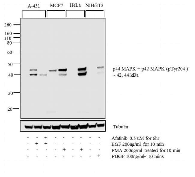
- WB analysis was performed on whole cell extracts (30 µg) of A-431 (1), A-431 treated with EGF (200 ng/mL for 10 minutes) (2), A-431 treated with Afatinib followed by EGF (0.5uM of Afatinib for 6hrs, 200 ng/mL for 10 minutes) (3), MCF7 (4) and MCF7 treated with PMA (200 ng/mL for 10 minutes) (5), HeLa (6) and HeLa treated with PMA (200 ng/mL for 10 minutes) (7), NIH/3T3 (8) and NIH/3T3 treated with PMA (200 ng/mL for 10 minutes) (9). The blot was probed with Phospho-ERK1/ERK2 (Tyr204) Rabbit Monoclonal Antibody (Product # MA5-15174, 1:1000 dilution) and detected by chemiluminescence using Goat anti-Rabbit IgG (H+L) Superclonal Secondary Antibody, HRP (Product # A27036, 0.25 µg/mL, 1:4000 dilution). 44, 42 kDa band corresponding to Phospho-ERK1/ERK2 (Tyr204) was detected and increased upon EGF and PMA treatment across cell lines and pre-treatment with Afatinib (antagonist) resulted in inhibition of Phospho-ERK1/ERK2 (Tyr204) in A-431 cell line upon EGF treatment. Protein samples were electrophoresed using Novex NuPAGE 4-12 % Bis-Tris gel (Product # NP0321BOX), XCell SureLock System (Product # EI0002) and Novex Sharp Pre-Stained Protein Standard (Product # LC5800). Resolved proteins were then wet transferred onto a nitrocellulose membrane. The membrane was probed with the relevant primary and secondary Antibody following blocking with 5 % skimmed milk. Chemiluminescent detection was performed using Pierce™ ECL Western Blotting Substrate (Product # 32106).

- Submitted by
- Invitrogen Antibodies (provider)
- Main image

- Experimental details
- WB analysis was performed on whole cell extracts (30 µg) of A-431 (1), A-431 treated with EGF (200 ng/mL for 10 minutes) (2), A-431 treated with Afatinib followed by EGF (0.5uM of Afatinib for 6hrs, 200 ng/mL for 10 minutes) (3), MCF7 (4) and MCF7 treated with PMA (200 ng/mL for 10 minutes) (5), HeLa (6) and HeLa treated with PMA (200 ng/mL for 10 minutes) (7), NIH/3T3 (8) and NIH/3T3 treated with PMA (200 ng/mL for 10 minutes) (9). The blot was probed with Phospho-ERK1/ERK2 (Tyr204) Rabbit Monoclonal Antibody (Product # MA5-15174, 1:1000 dilution) and detected by chemiluminescence using Goat anti-Rabbit IgG (H+L) Superclonal Secondary Antibody, HRP (Product # A27036, 0.25 µg/mL, 1:4000 dilution). 44, 42 kDa band corresponding to Phospho-ERK1/ERK2 (Tyr204) was detected and increased upon EGF and PMA treatment across cell lines and pre-treatment with Afatinib (antagonist) resulted in inhibition of Phospho-ERK1/ERK2 (Tyr204) in A-431 cell line upon EGF treatment. Protein samples were electrophoresed using Novex NuPAGE 4-12 % Bis-Tris gel (Product # NP0321BOX), XCell SureLock System (Product # EI0002) and Novex Sharp Pre-Stained Protein Standard (Product # LC5800). Resolved proteins were then wet transferred onto a nitrocellulose membrane. The membrane was probed with the relevant primary and secondary Antibody following blocking with 5 % skimmed milk. Chemiluminescent detection was performed using Pierce™ ECL Western Blotting Substrate (Product # 32106).
- Submitted by
- Invitrogen Antibodies (provider)
- Main image

- Experimental details
- Western blot analysis of extracts from NIH/3T3 cells treated with UV light and PDGF, using Phospho-p44/42 MAPK pErk1/2 pThr202/Tyr204 monoclonal antibody (Product # MA5-15174) (upper), Phospho-p38 MAPK (Thr180/Tyr182) monoclonal antibody (middle), and Phospho-SAPK/JNK (Thr183/Tyr185) monoclonal antibody (lower).

Supportive validation
- Submitted by
- Invitrogen Antibodies (provider)
- Main image

- Experimental details
- Immunofluorescent analysis of Phospho-p44/42 MAPK/Erk1/2 pThr202/Tyr204 in NIH/3T3 cells stimulated with Platelet-Derived Growth Factor, using a Phospho-p44/42 MAPK/Erk1/2 pThr202/Tyr204 monoclonal antibody (Product # MA5-15174) (green). Actin filaments are labeled with a fluorescent red phalloidin. DNA is labeled using a fluorescent blue dye.

Supportive validation
- Submitted by
- Invitrogen Antibodies (provider)
- Main image
- Experimental details
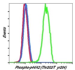
- Flow cytometric analysis of Phospho-p44/42 MAPK pErk1/2 pThr202/Tyr204 in U0126-inhibited (blue) or PMA-stimulated (green) Jurkat cells using a Phospho-p44/42 MAPK pErk1/2 pThr202/Tyr204 monoclonal antibody (Product # MA5-15174) compared to a nonspecific negative control antibody (red).

Supportive validation
- Submitted by
- Invitrogen Antibodies (provider)
- Main image
- Experimental details
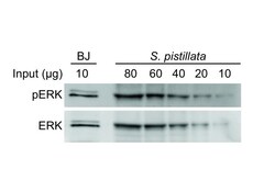
- Figure 4. Relative sensitivities of ERK antibodies toward the human and coral proteins. Immunoblot performed with anti-ERK and anti-phospho-ERK on total protein extracts of human fibroblasts (BJ) and Stylphora pistillata . The amount of protein loaded in each lane is indicated on the Supplementary Figure S4.

- Submitted by
- Invitrogen Antibodies (provider)
- Main image
- Experimental details
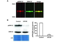
- Figure 2. Detection of ERK activity in corals. ( A ) Fluorescent immunoblot revealing activated (pERK) and total forms of ERK (ERK) present in Stylphora pistillata nubbins. Molecular weight standards in kilo Daltons (kDa) are indicated on the left side of the figure. ( B ) Immunoblot performed with ERK and pERK antibodies on protein extracts from coral nubbins incubated in the absence (Control) or presence of the MEK inhibitor U0126. Densitometric analysis of activated ERK intensities is presented on the right of the figure. The amido black total protein staining of the western blot membrane is shown as a loading control. The medians and standard deviations of three independent experiments are presented (***, p

- Submitted by
- Invitrogen Antibodies (provider)
- Main image
- Experimental details
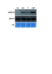
- Figure 3. Induction of Spi-ERK phosphorylation by thermal and UV stresses. Immunoblot performed with ERK and pERK antibodies on protein extracts from coral nubbins incubated for 30 minutes in control (Cont.), thermal stress (T), UV stress (UV) or a combination of thermal and UV stresses (UV + T) conditions. The amido black total protein staining of the western blot membrane is shown as a loading control.

- Submitted by
- Invitrogen Antibodies (provider)
- Main image

- Experimental details
- Figure 3 Left: extracellular signal regulated kinase (ERK)-phosphorylation upon reperfusion after reperfusion of kidneys with (controlled hyperthermia (CH)) or without (control) exposure to controlled hyperthermia during machine perfusion. Single values ( n = 3), mean and SEM are shown; * P < 0.05. Right: Protein expression of HSP 72 after reperfusion of kidneys with (CH) or without (control) exposure to controlled hyperthermia during machine perfusion.

- Submitted by
- Invitrogen Antibodies (provider)
- Main image

- Experimental details
- Figure 1 MEK1/2-RK1/2 and STAT3 play indispensable, yet distinct roles in mammary epithelial cell migration. A , both the STAT3 inhibitor S3I-201 and the MEK1/2 inhibitor U0126 blocked migration of the MCF-10A cells induced by EGF. Shown are percentage of wound healing (in 16 h) in the presence or the absence of EGF and/or the inhibitor. n = 3, ** p < 0.001; Student's t test. B , both S3I-201 and U0126 abrogated EGF-induced upregulation of CTEN expression. Data shown are obtained from samples with 8 h of treatment. C , Western blot showing the dynamic changes in the TNS3 and CTEN protein during 8 h of EGF treatment. Total and phosphorylated ERK1/2 and STAT3 were detected using specific antibodies. n = 3, * p < 0.05, ** p < 0.001; Student's t test. D , distinct binding profiles for STAT3 and ERK1/2 to the cten gene promoter at different time points of EGF treatment, graphed from the corresponding ChIP-PCR data. E , U0126 abrogated ERK1/2 phosphorylation at all time points but inhibited STAT3-Tyr705 phosphorylation only at late time points of EGF stimulation ( i.e. , 3 and 8 h). F , S3I-201 blocked STAT3-Tyr705 phosphorylation but had no effect on ERK1/2 phosphorylation. ChIP, chromatin immunoprecipitation; CTEN, C-terminal tension; EGF, epithelial growth factor; ERK1/2, extracellular signal-regulated protein kinase 1/2; MEK1/2, mitogen-activated protein kinase kinase 1/2; STAT3, signal transducer and activator of transcription 3; TNS3, tensin-3.

- Submitted by
- Invitrogen Antibodies (provider)
- Main image

- Experimental details
- Figure 3 The IL-6-STAT3-IL-6 feedback loop facilitates cell migration in response to continuous EGF treatment. A , IL-6 promoted EGF stimulated cell migration, whereas an anti-IL-6 antibody (alphaIL-6) significantly reduced cell migration. n = 3, * p < 0.05, Student's t test. B , IL-6 promoted STAT3-Y705 phosphorylation but had no effect on ERK1/2 phosphorylation. C , alphaIL-6 markedly reduced STAT3 phosphorylation but had no effect on ERK1/2 activation induced by EGF stimulation for 3 h. D and E , pharmacological inhibition of the EGFR, JAK, and Src kinases reduced or blocked STAT3 phosphorylation. F, relevant kinase inhibitors and anti-IL-6 antibody all decreased CTEN expression induced by EGF. G , nuclear translocation of STAT3 (STAT3-pY705) and ERK1/2 (pERK1/2) during continuous EGF stimulation. H , IL-6 transcription was regulated by ERK1/2 in the early phase ( e.g. , 10 min), but by STAT3 in the later phase ( e.g. , 3 h) of EGF stimulation. n = 3, * p < 0.05; ** p < 0.001, Student's t test. I , both S3I-201 and U0126 effectively blocked EGF-induced IL-6 expression in MCF-10A cells. EGF, epithelial growth factor; EGFR, epithelial growth factor receptor; ERK1/2, extracellular signal-regulated protein kinase 1/2; IL-6, interleukin-6; JAK, Janus kinase; STAT3, signal transducer and activator of transcription 3.
