Explore
 Validate
 Learn
Learn Western blot
 ELISA
ELISA Immunoprecipitation
Antibody data
- Antibody Data
- Antigen structure
- References [25]
- Comments [0]
- Validations
- Western blot [1]
- Immunocytochemistry [1]
- Other assay [13]
Submit
Validation data
Reference
Comment
Report error
- Product number
- 61-7400 - Provider product page

- Provider
- Invitrogen Antibodies
- Product name
- ERK1/ERK2 Polyclonal Antibody
- Antibody type
- Polyclonal
- Antigen
- Synthetic peptide
- Reactivity
- Human, Mouse, Rat, Rabbit
- Host
- Rabbit
- Isotype
- IgG
- Vial size
- 100 µg
- Concentration
- 0.25 mg/mL
- Storage
- -20°C
Submitted references Empagliflozin mitigates type 2 diabetes-associated peripheral neuropathy: a glucose-independent effect through AMPK signaling.
Regulation of Sirtuin-3 and ERK1/2/p38MAPK by the combination Ga nanoparticles/γ-radiation low dosage: an effective approach for treatment of hepatocellular carcinoma.
Acylated Ghrelin and The Regulation of Lipid Metabolism in The Intestine.
The role of fibroblast growth factor signalling in Echinococcus multilocularis development and host-parasite interaction.
Cardiac hypertrophy in mice submitted to a swimming protocol: influence of training volume and intensity on myocardial renin-angiotensin system.
CAPN1 is a novel binding partner and regulator of the tumor suppressor NF1 in melanoma.
Extracellular Signal-Regulated Kinase Signaling in CD4-Expressing Cells Inhibits Osteochondromas.
Downregulation of Lnc-Spry1 mediates TGF-β-induced epithelial-mesenchymal transition by transcriptional and posttranscriptional regulatory mechanisms.
Iterative Modeling Reveals Evidence of Sequential Transcriptional Control Mechanisms.
Analysis of the V2 Vasopressin Receptor (V2R) Mutations Causing Partial Nephrogenic Diabetes Insipidus Highlights a Sustainable Signaling by a Non-peptide V2R Agonist.
Targeting SALL4 by entinostat in lung cancer.
Transgenic mice with increased astrocyte expression of IL-6 show altered effects of acute ethanol on synaptic function.
PARP targeting counteracts gliomagenesis through induction of mitotic catastrophe and aggravation of deficiency in homologous recombination in PTEN-mutant glioma.
CCL2-ethanol interactions and hippocampal synaptic protein expression in a transgenic mouse model.
Increased astrocyte expression of IL-6 or CCL2 in transgenic mice alters levels of hippocampal and cerebellar proteins.
GPR30 activation is neither necessary nor sufficient for acute neuroprotection by 17β-estradiol after an ischemic injury in organotypic hippocampal slice cultures.
Brain-derived neurotrophic factor protects against cardiac dysfunction after myocardial infarction via a central nervous system-mediated pathway.
Neuroadaptive changes in cerebellar neurons induced by chronic exposure to IL-6.
Id-1 activates Akt-mediated Wnt signaling and p27(Kip1) phosphorylation through PTEN inhibition.
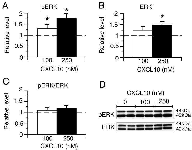
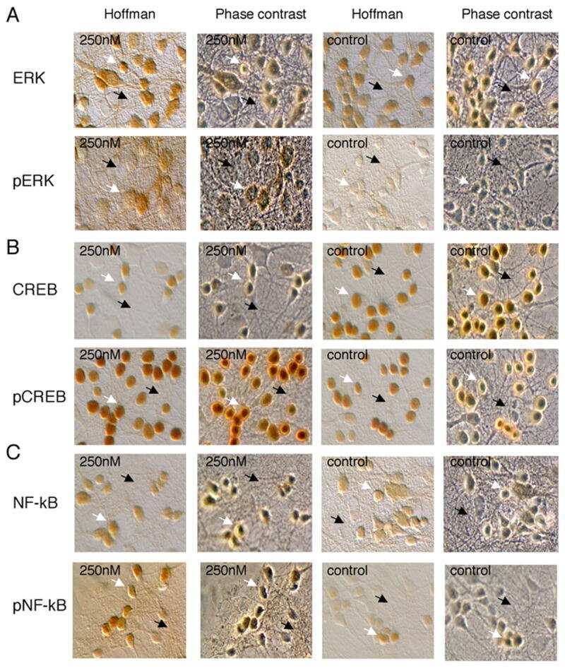
Chronic CXCL10 alters the level of activated ERK1/2 and transcriptional factors CREB and NF-kappaB in hippocampal neuronal cell culture.
Single and combined silencing of ERK1 and ERK2 reveals their positive contribution to growth signaling depending on their expression levels.
Inhibitor of DNA binding 1 activates vascular endothelial growth factor through enhancing the stability and activity of hypoxia-inducible factor-1alpha.
In situ analysis of integrin and growth factor receptor signaling pathways in human glioblastomas suggests overlapping relationships with focal adhesion kinase activation.
A role for mitogen-activated protein kinase(Erk1/2) activation and non-selective pore formation in P2X7 receptor-mediated thymocyte death.
Conditional, tissue-specific expression of Q205L Galphai2 in vivo mimics insulin activation of c-Jun N-terminal kinase and p38 kinase.
Abdelkader NF, Elbaset MA, Moustafa PE, Ibrahim SM
Archives of pharmacal research 2022 Jul;45(7):475-493
Archives of pharmacal research 2022 Jul;45(7):475-493
Regulation of Sirtuin-3 and ERK1/2/p38MAPK by the combination Ga nanoparticles/γ-radiation low dosage: an effective approach for treatment of hepatocellular carcinoma.
Abdalla MS, El-Mahdy EM, Mansour SZ, Elsonbaty SM, Amin MH
Journal, genetic engineering & biotechnology 2022 Jul 1;20(1):93
Journal, genetic engineering & biotechnology 2022 Jul 1;20(1):93
Acylated Ghrelin and The Regulation of Lipid Metabolism in The Intestine.
Auclair N, Patey N, Melbouci L, Ou Y, Magri-Tomaz L, Sané A, Garofalo C, Levy E, St-Pierre DH
Scientific reports 2019 Nov 29;9(1):17975
Scientific reports 2019 Nov 29;9(1):17975
The role of fibroblast growth factor signalling in Echinococcus multilocularis development and host-parasite interaction.
Förster S, Koziol U, Schäfer T, Duvoisin R, Cailliau K, Vanderstraete M, Dissous C, Brehm K
PLoS neglected tropical diseases 2019 Mar;13(3):e0006959
PLoS neglected tropical diseases 2019 Mar;13(3):e0006959
Cardiac hypertrophy in mice submitted to a swimming protocol: influence of training volume and intensity on myocardial renin-angiotensin system.
Soares DDS, Pinto GH, Lopes A, Caetano DSL, Nascimento TG, Andrades ME, Clausell N, Rohde LEP, Leitão SAT, Biolo A
American journal of physiology. Regulatory, integrative and comparative physiology 2019 Jun 1;316(6):R776-R782
American journal of physiology. Regulatory, integrative and comparative physiology 2019 Jun 1;316(6):R776-R782
CAPN1 is a novel binding partner and regulator of the tumor suppressor NF1 in melanoma.
Alon M, Arafeh R, Lee JS, Madan S, Kalaora S, Nagler A, Abgarian T, Greenberg P, Ruppin E, Samuels Y
Oncotarget 2018 Jul 27;9(58):31264-31277
Oncotarget 2018 Jul 27;9(58):31264-31277
Extracellular Signal-Regulated Kinase Signaling in CD4-Expressing Cells Inhibits Osteochondromas.
Wehenkel M, Corr M, Guy CS, Edwards BA, Castellaw AH, Calabrese C, Pagès G, Pouysségur J, Vogel P, McGargill MA
Frontiers in immunology 2017;8:482
Frontiers in immunology 2017;8:482
Downregulation of Lnc-Spry1 mediates TGF-β-induced epithelial-mesenchymal transition by transcriptional and posttranscriptional regulatory mechanisms.
Rodríguez-Mateo C, Torres B, Gutiérrez G, Pintor-Toro JA
Cell death and differentiation 2017 May;24(5):785-797
Cell death and differentiation 2017 May;24(5):785-797
Iterative Modeling Reveals Evidence of Sequential Transcriptional Control Mechanisms.
Cheng CS, Behar MS, Suryawanshi GW, Feldman KE, Spreafico R, Hoffmann A
Cell systems 2017 Mar 22;4(3):330-343.e5
Cell systems 2017 Mar 22;4(3):330-343.e5
Analysis of the V2 Vasopressin Receptor (V2R) Mutations Causing Partial Nephrogenic Diabetes Insipidus Highlights a Sustainable Signaling by a Non-peptide V2R Agonist.
Makita N, Sato T, Yajima-Shoji Y, Sato J, Manaka K, Eda-Hashimoto M, Ootaki M, Matsumoto N, Nangaku M, Iiri T
The Journal of biological chemistry 2016 Oct 21;291(43):22460-22471
The Journal of biological chemistry 2016 Oct 21;291(43):22460-22471
Targeting SALL4 by entinostat in lung cancer.
Yong KJ, Li A, Ou WB, Hong CK, Zhao W, Wang F, Tatetsu H, Yan B, Qi L, Fletcher JA, Yang H, Soo R, Tenen DG, Chai L
Oncotarget 2016 Nov 15;7(46):75425-75440
Oncotarget 2016 Nov 15;7(46):75425-75440
Transgenic mice with increased astrocyte expression of IL-6 show altered effects of acute ethanol on synaptic function.
Hernandez RV, Puro AC, Manos JC, Huitron-Resendiz S, Reyes KC, Liu K, Vo K, Roberts AJ, Gruol DL
Neuropharmacology 2016 Apr;103:27-43
Neuropharmacology 2016 Apr;103:27-43
PARP targeting counteracts gliomagenesis through induction of mitotic catastrophe and aggravation of deficiency in homologous recombination in PTEN-mutant glioma.
Majuelos-Melguizo J, Rodríguez MI, López-Jiménez L, Rodríguez-Vargas JM, Martí Martín-Consuegra JM, Serrano-Sáenz S, Gavard J, de Almodóvar JM, Oliver FJ
Oncotarget 2015 Mar 10;6(7):4790-803
Oncotarget 2015 Mar 10;6(7):4790-803
CCL2-ethanol interactions and hippocampal synaptic protein expression in a transgenic mouse model.
Gruol DL, Vo K, Bray JG, Roberts AJ
Frontiers in integrative neuroscience 2014;8:29
Frontiers in integrative neuroscience 2014;8:29
Increased astrocyte expression of IL-6 or CCL2 in transgenic mice alters levels of hippocampal and cerebellar proteins.
Gruol DL, Vo K, Bray JG
Frontiers in cellular neuroscience 2014;8:234
Frontiers in cellular neuroscience 2014;8:234
GPR30 activation is neither necessary nor sufficient for acute neuroprotection by 17β-estradiol after an ischemic injury in organotypic hippocampal slice cultures.
Lamprecht MR, Morrison B 3rd
Brain research 2014 May 14;1563:131-7
Brain research 2014 May 14;1563:131-7
Brain-derived neurotrophic factor protects against cardiac dysfunction after myocardial infarction via a central nervous system-mediated pathway.
Okada S, Yokoyama M, Toko H, Tateno K, Moriya J, Shimizu I, Nojima A, Ito T, Yoshida Y, Kobayashi Y, Katagiri H, Minamino T, Komuro I
Arteriosclerosis, thrombosis, and vascular biology 2012 Aug;32(8):1902-9
Arteriosclerosis, thrombosis, and vascular biology 2012 Aug;32(8):1902-9
Neuroadaptive changes in cerebellar neurons induced by chronic exposure to IL-6.
Gruol DL, Puro A, Hao C, Blakely P, Janneke E, Vo K
Journal of neuroimmunology 2011 Oct 28;239(1-2):28-36
Journal of neuroimmunology 2011 Oct 28;239(1-2):28-36
Id-1 activates Akt-mediated Wnt signaling and p27(Kip1) phosphorylation through PTEN inhibition.
Lee JY, Kang MB, Jang SH, Qian T, Kim HJ, Kim CH, Kim Y, Kong G
Oncogene 2009 Feb 12;28(6):824-31
Oncogene 2009 Feb 12;28(6):824-31
Chronic CXCL10 alters the level of activated ERK1/2 and transcriptional factors CREB and NF-kappaB in hippocampal neuronal cell culture.
Bajova H, Nelson TE, Gruol DL
Journal of neuroimmunology 2008 Mar;195(1-2):36-46
Journal of neuroimmunology 2008 Mar;195(1-2):36-46
Single and combined silencing of ERK1 and ERK2 reveals their positive contribution to growth signaling depending on their expression levels.
Lefloch R, Pouysségur J, Lenormand P
Molecular and cellular biology 2008 Jan;28(1):511-27
Molecular and cellular biology 2008 Jan;28(1):511-27
Inhibitor of DNA binding 1 activates vascular endothelial growth factor through enhancing the stability and activity of hypoxia-inducible factor-1alpha.
Kim HJ, Chung H, Yoo YG, Kim H, Lee JY, Lee MO, Kong G
Molecular cancer research : MCR 2007 Apr;5(4):321-9
Molecular cancer research : MCR 2007 Apr;5(4):321-9
In situ analysis of integrin and growth factor receptor signaling pathways in human glioblastomas suggests overlapping relationships with focal adhesion kinase activation.
Riemenschneider MJ, Mueller W, Betensky RA, Mohapatra G, Louis DN
The American journal of pathology 2005 Nov;167(5):1379-87
The American journal of pathology 2005 Nov;167(5):1379-87
A role for mitogen-activated protein kinase(Erk1/2) activation and non-selective pore formation in P2X7 receptor-mediated thymocyte death.
Auger R, Motta I, Benihoud K, Ojcius DM, Kanellopoulos JM
The Journal of biological chemistry 2005 Jul 29;280(30):28142-51
The Journal of biological chemistry 2005 Jul 29;280(30):28142-51
Conditional, tissue-specific expression of Q205L Galphai2 in vivo mimics insulin activation of c-Jun N-terminal kinase and p38 kinase.
Guo JH, Wang HY, Malbon CC
The Journal of biological chemistry 1998 Jun 26;273(26):16487-93
The Journal of biological chemistry 1998 Jun 26;273(26):16487-93
No comments: Submit comment
Supportive validation
- Submitted by
- Invitrogen Antibodies (provider)
- Main image
- Experimental details
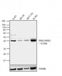
- Western blot analysis was performed on whole cell extracts (30 µg lysate) of A-431 (Lane 1), HT-29 (Lane 2), HCT 116 (Lane 3) and K-562 (Lane 4). The blot was probed with Rabbit Anti-ERK1/ERK2 Polyclonal Antibody (Product # 61-7400, 1:1,000 dilution) and detected by chemiluminescence using Goat anti-Rabbit IgG (H+L) Superclonal™ Secondary Antibody, HRP conjugate (Product # A27036, 0.4 µg/mL, 1:2500 dilution). A 42 kDa band corresponding to ERK1 was observed across the cell lines tested. Known quantity of protein samples were electrophoresed using Novex® NuPAGE® 4-12 % Bis-Tris gel (Product # NP0321BOX), XCell SureLock™ Electrophoresis System (Product # EI0002) and Novex® Sharp Pre-Stained Protein Standard (Product # LC5800). Resolved proteins were then transferred onto a nitrocellulose membrane with iBlot® 2 Dry Blotting System (Product # IB21001). The membrane was probed with the relevant primary and secondary Antibody following blocking with 5 % skimmed milk. Chemiluminescent detection was performed using Pierce™ ECL Western Blotting Substrate (Product # 32106).

Supportive validation
- Submitted by
- Invitrogen Antibodies (provider)
- Main image
- Experimental details
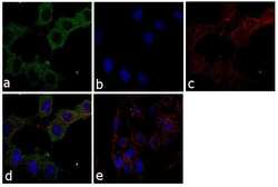
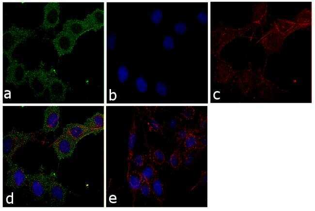
- Immunofluorescence analysis of MAPKINASE (ERK1+ERK2) was performed using 70% confluent log phase RSC96 cells. The cells were fixed with 4% paraformaldehyde for 10 minutes, permeabilized with 0.1% Triton™ X-100 for 10 minutes, and blocked with 1% BSA for 1 hour at room temperature. The cells were labeled with ERK1/ERK2 Rabbit Polyclonal Antibody (Product # 61-7400) at 2µg/mL in 0.1% BSA and incubated for 3 hours at room temperature and then labeled with Goat anti-Rabbit IgG (H+L) Superclonal™ Secondary Antibody, Alexa Fluor® 488 conjugate (Product # A27034) at a dilution of 1:2000 for 45 minutes at room temperature (Panel a: green). Nuclei (Panel b: blue) were stained with SlowFade® Gold Antifade Mountant with DAPI (Product # S36938). F-actin (Panel c: red) was stained with Rhodamine Phalloidin (Product # R415, 1:300). Panel d represents the merged image showing cytoplasmic localization. Panel e shows the no primary antibody control. The images were captured at 60X magnification.
Supportive validation
- Submitted by
- Invitrogen Antibodies (provider)
- Main image

- Experimental details
- NULL

- Submitted by
- Invitrogen Antibodies (provider)
- Main image

- Experimental details
- NULL
- Submitted by
- Invitrogen Antibodies (provider)
- Main image

- Experimental details
- NULL
- Submitted by
- Invitrogen Antibodies (provider)
- Main image
- Experimental details
- NULL

- Submitted by
- Invitrogen Antibodies (provider)
- Main image

- Experimental details
- NULL

- Submitted by
- Invitrogen Antibodies (provider)
- Main image
- Experimental details
- NULL
- Submitted by
- Invitrogen Antibodies (provider)
- Main image

- Experimental details
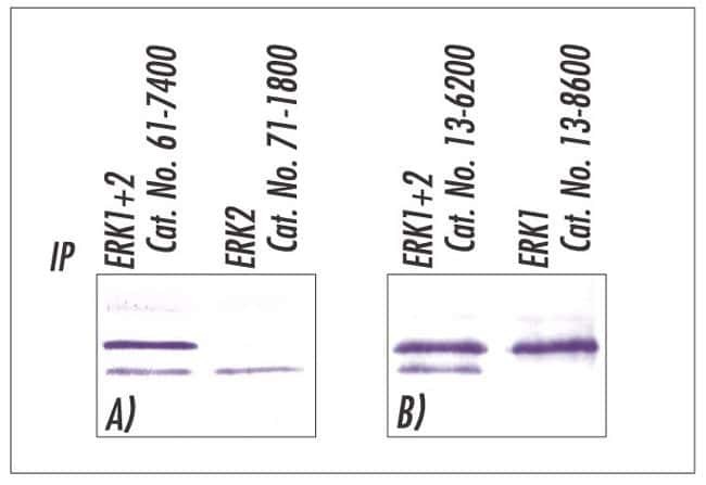
- Western blot analysis following immunoprecipitation of mouse brain homogenates using Zymed's ERK1+ERK2 (MAP kinase) (Product # 61-7400, 13-6200), ERK1 (Product # 13-8600), and ERK2 (Product # 71-1800) antibodies.
- Submitted by
- Invitrogen Antibodies (provider)
- Main image

- Experimental details
- Figure 4 ERK2 is expressed in the majority of chondrocytes in DKO CD4 mice . (A) Chondrocytes were isolated from individual WT, Erk1 -/- .Erk2 fl/fl .CD4cre - (E1), or DKO CD4 mice and cultured in vitro . DNA was isolated and analyzed by PCR for the presence of the wild-type, floxed, or excised Erk2 allele. Tail DNA is shown as controls for the wild-type and floxed alleles, while DNA from thymocytes serves as the positive control for the excised allele. (B) Cultured chondrocytes were lysed and analyzed by western blot for ERK1, ERK2, type II collagen (Col2A1), and beta-actin. (C) Planar X-ray (left) and surgical microscopy images of a representative lesion from a DKO CD4 mouse. (D) Three microdissected tumors from individual mice greater than 20 weeks of age were analyzed by PCR to determine whether the Erk2 gene was wild-type, floxed, or excised by Cre recombinase. DNA from DKO CD4 thymocytes is shown as a positive control for Erk2 excision, while tail DNA is a negative control for excision. The first row contains primers that amplify the wild-type, floxed, or excised alleles, the second row is primers that only amplify the excised allele. The third row contains primers that amplify the wild-type or floxed alleles.

- Submitted by
- Invitrogen Antibodies (provider)
- Main image
- Experimental details
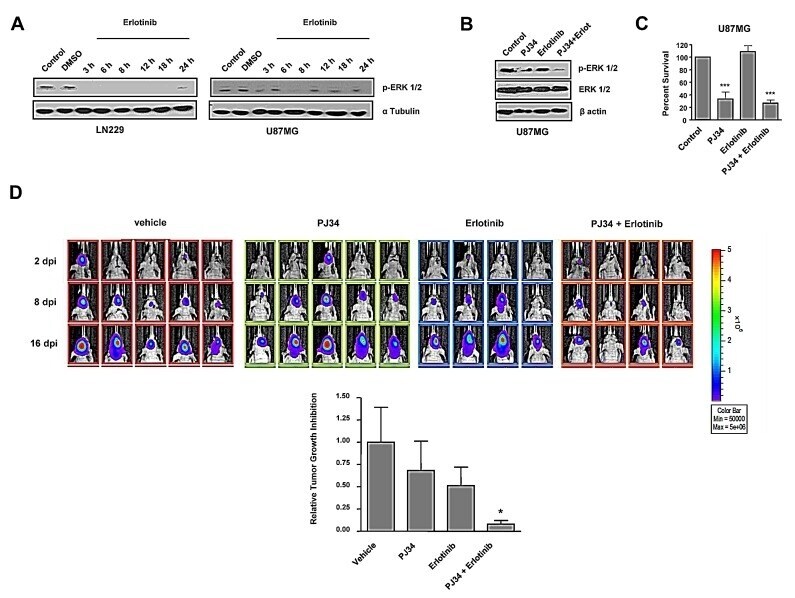
- Figure 4 In vitro effect of EGFR inhibitor erlotinib and decreased tumours growth in vivo after combined treatment with PARP inhibitor and erlotinib A. Western blot analysis of p-ERK-1/2 expression levels at different times following erlotinib treatment.. B,C. B,C. U87MG cells were treated with erlotinib alone or combined with PJ34 during 72 hours. (B) p-ERK-1/2 expression was measured by Western Blot. (C) MTT reduction was analysed. *** p < 0.001 versus control group by t-test. D. Mice were inoculated with U87MG-luciferase human cell line. Localization and intensity of luciferase expression were monitored by in vivo bioluminiscence imaging (dpi, days post cells injection). Representation of tumours growth inhibition on the 16 th day. A statistically significant reduction is observed in the combined treatment of PJ34 and erlotinib. * p < 0.05 versus control group by t-test. Data are represented as mean +- SEM of 3 independent experiments.
- Submitted by
- Invitrogen Antibodies (provider)
- Main image
- Experimental details
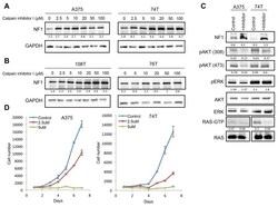
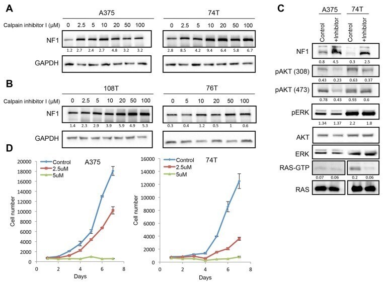
- Figure 3 CAPN1 inhibition stabilizes NF1 levels, affecting RAS signaling and cell proliferation (A) NF1 wild type cells (A375, 74T) were treated with increasing concentrations of Calpain inhibitor I (muM) for 6 hours or with DMSO as control, and NF1 levels were tested by immunoblot. (B) NF1 mutant cells (108T, 76T) were treated with increasing concentrations of Calpain inhibitor I (muM) for 6 hours or with DMSO as control, and NF1 levels were tested by immunoblot. Ratios of NF1 levels to GAPDH were generated using Image lab (BioRad) and Microsoft Excel analysis. (C) 74T and A375 cells were treated with 3 muM or 5 muM of Calpain inhibitor I for 16 hours, respectively. Cell lysates were analyzed by western blot with the indicated antibodies. RAS-GTP levels were assessed by RAS pulldown assay after treatment with 50 muM of Calpain inhibitor I, respectively for 6 hours. (D) A375 and 74T cells were seeded in the 96 well plates with 10% FBS and cells were treatedwith 2.5 or 5 muM of Calpain inhibitor I, respectively. DMSO was used as a control for this experiment. The average cell number was measured by assessing DNA content using SYBR green I in two independent experiments with six replicates each. Error bars, s.e.m.
- Submitted by
- Invitrogen Antibodies (provider)
- Main image
- Experimental details
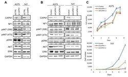
- Figure 4 Suppression of CAPN1 by shRNA stabilizes NF1 levels and affects Ras signaling and cell proliferation (A) Immunoblot of lysates generated from siRNA mediated CAPN1 knockdown and control tested by transient transfection of 100 nM for 72 hours. (B) Immunoblots of lysates generated from two shRNA mediated CAPN1 knockdown (shCAPN1-1 and shCAPN1-2) compared to the control vector. Quantification values are given under the blots generated by Image lab (BioRad) and Microsoft Excel analysis. (C) 74T and A375 cells stably expressing shRNA against CAPN1 (shCAPN1-1 or shCAPN1-2) were grown in 96 well plates with 10% or 2.5% FBS, respectively. The average cell number was measured by assessing DNA content using SYBR green I in two independent experiments with six replicates each. Error bars, s.e.m.

- Submitted by
- Invitrogen Antibodies (provider)
- Main image
- Experimental details
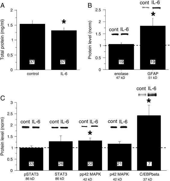
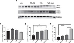
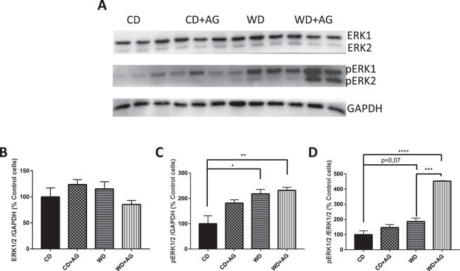
- Figure 5 Effect of acylated ghrelin on ERK1/ERK2 activation in the jejunum. Syrian Golden Hamsters were submitted to conventional chow (CD) or Western (WD) diets for 8 weeks. For the last 2 weeks of treatment, animals were continuously administered with acylated ghrelin (AG, 100 nmol/kg/day) or physiological saline. Before sacrifice, hamsters were fasted for 16 h before receiving an olive oil gavage (200 uL). Jejunum samples were homogenized in a lysis buffer and proteins lysates were analyzed by Western Blot with ERK1/ERK2 ( A,B ) and phosphoERK1/ERK2 ( A,C ) antibodies as indicated. The ratio of pERK1/2/ERK1/2 was also analyzed. ( D ) For accurate normalization, the same blot was probed with GAPDH. A representative blot is shown, illustrating an experiment in triplicate on the same gel and at the same time exposure. The results of every experiment are shown as mean +- SEM of n = 4-6 animals * P < 0.05; ** P < 0.01; *** P < 0.001; **** P < 0.0001.
- Submitted by
- Invitrogen Antibodies (provider)
- Main image

- Experimental details
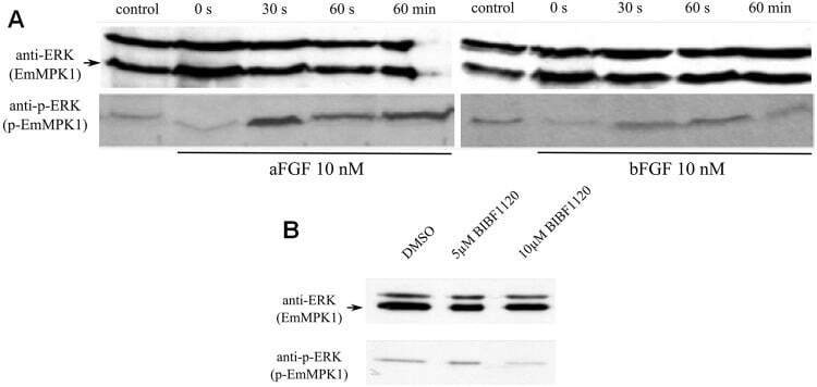
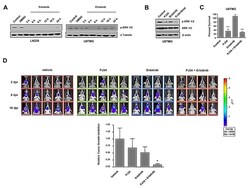
- Fig 7 Effects of host FGFs and BIBF 1120 on EmMPK1 phosphorylation in metacestode vesicles. A , axenically cultivated metacestode vesicles were incubated in cMEM (control) or in medium without FCS (0 s), upon which FGF1 (aFGF) or FGF2 (bFGF) were added at a concentration of 10 nM for 30 sec (30s), 60 sec (60s) or 60 min (60min). Protein lysates were subsequently separated by 12% SDS-PAGE and Western blots were analysed using polyclonal antibodies against Erk-like MAP kinases (anti-ERK) or double phosphorylated Erk-like MAP kinases (anti-p-ERK). B , axenically cultivated metacestode vesicles were incubated with DMSO (negative control), 5 mM or 10 mM BIBF1120 (30 min each) and cell lysates were subsequently analysed as described above. Both experiments were performed in triplicate with similar results.