 Explore
 Validate
 Learn
Learn Western blot
Antibody data
- Antibody Data
- Antigen structure
- References [0]
- Comments [0]
- Validations
- Western blot [3]
- Immunocytochemistry [1]
- Immunohistochemistry [1]
Submit
Validation data
Reference
Comment
Report error
- Product number
- PA5-36776 - Provider product page

- Provider
- Invitrogen Antibodies
- Product name
- Phospho-ERK1/ERK2 (Tyr204) Polyclonal Antibody
- Antibody type
- Polyclonal
- Antigen
- Synthetic peptide
- Description
- This antibody detects endogenous protein at a molecular weight of 42 and 44 kDa. Purity is >95% by SDS-PAGE.
- Reactivity
- Human, Mouse, Rat
- Host
- Rabbit
- Isotype
- IgG
- Vial size
- 100 µL
- Concentration
- 1 mg/mL
- Storage
- Store at 4°C short term. For long term storage, store at -20°C, avoiding freeze/thaw cycles.
No comments: Submit comment
Supportive validation
- Submitted by
- Invitrogen Antibodies (provider)
- Main image

- Experimental details
- Western blot analysis of Phospho-ERK1/ERK2 (Tyr204) in Lane 1: L02 starved for 16 hours whole cell lysate (40 µg), Lane 2: L02 starved for 16 hours then treated with LPS (1000 ng/mL) for 15 minutes whole cell lysate, Lane 3: L02 starved for 16 hours then treated with LPS (1000 ng/mL) for 30 minutes whole cell lysate. Samples were incubated with Phospho-ERK1/ERK2 (Tyr204) polyclonal antibody (Product # PA5-36776) at a dilution of 1:500.

- Submitted by
- Invitrogen Antibodies (provider)
- Main image

- Experimental details
- Western blot analysis of Phospho-ERK1/ERK2 (Tyr204) in Lane 1: HEK293T whole cell lysate treated with PMA (100 nM,15mins), Lane 2: sp2/0 whole cell lysate treated with PMA (100 nM,15mins), Lane 3: PC12 whole cell lysate treated with PMA (100 nM,15mins). Samples were incubated with Phospho-ERK1/ERK2 (Tyr204) polyclonal antibody (Product # PA5-36776) at a dilution of 1:500.

- Submitted by
- Invitrogen Antibodies (provider)
- Main image

- Experimental details
- Western blot analysis of Phospho-ERK1+ERK2 pTyr204 using Phospho-ERK1+ERK2 pTyr204 polyclonal antibody (Product # PA5-36776) at a dilution of 1:500. Lane 1: HEK293T whole cell lysate treated with PMA (100nM, 15min), Lane 2: sp2/0 whole cell lysate treated with PMA (100nM, 15min), Lane 3: PC12 whole cell lysate treated with PMA (100nM, 15min).

Supportive validation
- Submitted by
- Invitrogen Antibodies (provider)
- Main image
- Experimental details
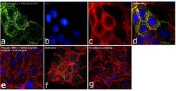
- Immunofluorescence analysis of Phospho-ERK1/ERK2 (Tyr204) was performed using 90% confluent log phase A-431 cells treated with 200 ng/mL of EGF for 10 minutes. The cells were fixed with 4% paraformaldehyde for 10 minutes, permeabilized with 0.1% Triton™ X-100 for 15 min, and blocked with 1% BSA for 1 hour at room temperature. The cells were labeled with Phospho-ERK1/ERK2 (Tyr204) Rabbit Polyclonal Antibody (Product # PA5-36776) at 5 µg in 0.1% BSA and incubated overnight at 4 degree Celsius and then labelled with Goat anti-Rabbit IgG (H+L) Superclonal™ Secondary Antibody, Alexa Fluor® 488 conjugate (Product # A27034) at a dilution of 1:2000 for 45 minutes at room temperature (Panel a: green). Nuclei (Panel b: blue) were stained with SlowFade® Gold Antifade Mountant with DAPI (Product # S36938). F-actin (Panel c: red) was stained with Rhodamine Phalloidin (Product # R415, 1:100). Panel d represents the merged image showing membrane localization. Panel e represents cells treated with antagonist, Afatinib (1µM for 6hrs) followed by EGF (200 ng/mL for 10 minutes), showing no signal. Panel f shows untreated cells with nuclear staining. Panel g represents control cells with no primary antibody to assess background. The images were captured at 60X magnification.

Supportive validation
- Submitted by
- Invitrogen Antibodies (provider)
- Main image

- Experimental details
- Immunohistochemical analysis of Phospho-ERK1+ERK2 pTyr204 in paraffin-embedded human breast carcinoma using Phospho-ERK1+ERK2 pTyr204 polyclonal antibody (Product # PA5-36776) at a dilution of 1:100.
