 Explore
 Validate
 Learn
Learn Western blot
Antibody data
- Antibody Data
- Antigen structure
- References [0]
- Comments [0]
- Validations
- Western blot [3]
- Immunocytochemistry [1]
- Immunohistochemistry [1]
- Flow cytometry [1]
- Other assay [2]
Submit
Validation data
Reference
Comment
Report error
- Product number
- MA5-15605 - Provider product page

- Provider
- Invitrogen Antibodies
- Product name
- ERK1/ERK2 Monoclonal Antibody (3F8B3)
- Antibody type
- Monoclonal
- Antigen
- Purifed from natural sources
- Description
- MA5-15605 targets p44/42 MAPK (Erk1/2) in FACS, IHC, WB and IF applications and shows reactivity with Human and mouse samples.
- Reactivity
- Human, Mouse, Rat
- Host
- Mouse
- Isotype
- IgG
- Antibody clone number
- 3F8B3
- Vial size
- 100 µg
- Concentration
- 1 mg/mL
- Storage
- Store at 4°C short term. For long term storage, store at -20°C, avoiding freeze/thaw cycles.
No comments: Submit comment
Supportive validation
- Submitted by
- Invitrogen Antibodies (provider)
- Main image

- Experimental details
- Western blot analysis of p42, 44 ERK (1, 2) was performed by loading 20 µg of the indicated whole cell lysates and 5 µl of PageRuler Plus Prestained Protein Ladder (Product # 26619) per well onto a 4-20% Tris-Glycine polyacrylamide gel (Product # WT4202BX10). Proteins were transferred to a nitrocellulose membrane using the G2 Blotter (Product # 62288), and blocked with 5% Milk in TBST for 1 hour at room temperature. p42, 44 ERK (1, 2) was detected at 42kDa using a p42, 44 ERK (1, 2) mouse monoclonal antibody (Product # MA5-15605) at a dilution of 1:1000 in blocking buffer for 1 hour at room temperature on a rocking platform, followed by a Goat anti-Mouse IgG (H+L) Superclonal™ Secondary Antibody, HRP conjugate (Product # A28177) at a dilution of 1:1000 for at least 30 minutes at room temperature. Chemiluminescent detection was performed using SuperSignal West Pico (Product # 34078).

- Submitted by
- Invitrogen Antibodies (provider)
- Main image

- Experimental details
- Knockdown of ERK1/ERK2 was achieved by transfecting A549 cells with ERK1/ERK2 specific validated siRNAs (Silencer® select Product # s11140, s11138 ). Western blot analysis (Fig. a) was performed using whole cell extracts from the ERK1/ERK2 knockdown cells (lane 3), non-specific scrambled siRNA transfected cells (lane 2) and untransfected cells (lane 1). The blot was probed with ERK1/ERK2 Monoclonal Antibody (Product # MA5-15605, 1:1000 dilution) and Goat anti-Mouse IgG (H+L) Superclonal™ Secondary Antibody, HRP conjugate (Product # A28177, 0.25 µg/mL, 1:4000 dilution). Densitometric analysis of this western blot is shown in histogram (Fig. b). Decrease in signal upon siRNA mediated knock down confirms that antibody is specific to GAPDH.

- Submitted by
- Invitrogen Antibodies (provider)
- Main image

- Experimental details
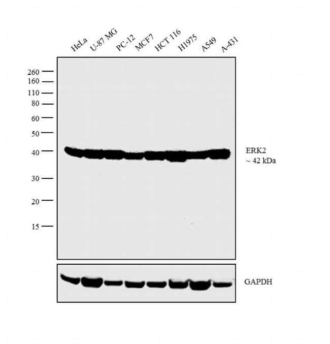
- Western blot analysis was performed on whole cell extracts (30 µg lysate) of HeLa (Lane 1), U-87 MG (Lane 2), PC-12 (Lane 3), MCF7 (Lane 4), HCT 116 (Lane 5), H1975 (Lane 6), A549 (Lane 7) and A-431 (Lane 8 ). The blot was probed with Anti-ERK1/ERK2 Monoclonal Antibody (Product # MA5-15605, 1:1000 dilution) and detected by chemiluminescence using Goat anti-Mouse IgG (H+L) Superclonal™ Secondary Antibody, HRP conjugate (Product # A28177, 0.25 µg/mL, 1:4000 dilution). A 42 kDa band corresponding to ERK2 was observed across the cell lines tested.
Supportive validation
- Submitted by
- Invitrogen Antibodies (provider)
- Main image
- Experimental details
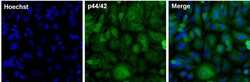
- Immunofluorescent analysis of p44, 42 MAPK (Erk1/2) in HeLa cells. The cells were fixed with 4% paraformaldehyde in PBS for 15 minutes at room temperature, permeabilized with 0.1% Triton X-100 for 15 minutes, and blocked with 3% BSA for 30 minutes at room temperature. Cells were stained with a p44/42 MAPK (Erk1/2) mouse monoclonal antibody (Product # MA5-15605) at a dilution of 1:250 in blocking buffer for 1 hour at room temperature, and then incubated with a Goat anti-Mouse IgG (H+L) Superclonal™ Secondary Antibody, Alexa Fluor® 488 conjugate (Product # A28175) at a dilution of 1:1000 for at least 30 minutes at a room temperature in the dark (green). Nuclei (blue) were stained with Hoechst 33342 (Product # 62249). Images were taken on a Thermo Scientific ToxInsight Instrument at 20X magnification.

Supportive validation
- Submitted by
- Invitrogen Antibodies (provider)
- Main image
- Experimental details
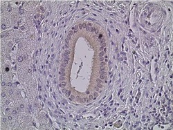
- Immunohistochemistry was performed on human liver tissue. Tissue was deparaffinized with xylene, followed by rehydration in sequential washes of 100% ethanol, 95% ethanol, 80% ethanol, and water. To expose target proteins, antigen retrieval was performed using 10mM sodium citrate (pH 6.0) and heated for 20 min. Following antigen retrieval, tissues were blocked in a 10% goat serum (Product # 31872) in wash buffer solution for 30-60 minutes at room temperature. Tissue was then probed with a p42, 44 ERK (1, 2) Mouse Monoclonal antibody (Product # MA5-15605) at a dilution of 1:100 in 10% goat serum in wash buffer overnight at 4°C in a humidified chamber. Negative control tissue received no primary antibody. Tissues were washed extensively with PBST, and detection was performed using a goat anti-mouse IgG-HRP secondary antibody at a dilution of 1:500 followed by colorimetric detection using metal enhanced DAB. Tissues were then counterstained with hematoxylin and prepped for mounting and imaging.

Supportive validation
- Submitted by
- Invitrogen Antibodies (provider)
- Main image

- Experimental details
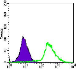
- Flow cytometric analysis of Jurkat cells using p44+42 MAPK (Erk1, 2) monoclonal antibody (Product # MA5-15605) (green) and negative control (purple).
Supportive validation
- Submitted by
- Invitrogen Antibodies (provider)
- Main image
- Experimental details
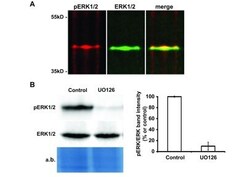
- Figure 2. Detection of ERK activity in corals. ( A ) Fluorescent immunoblot revealing activated (pERK) and total forms of ERK (ERK) present in Stylphora pistillata nubbins. Molecular weight standards in kilo Daltons (kDa) are indicated on the left side of the figure. ( B ) Immunoblot performed with ERK and pERK antibodies on protein extracts from coral nubbins incubated in the absence (Control) or presence of the MEK inhibitor U0126. Densitometric analysis of activated ERK intensities is presented on the right of the figure. The amido black total protein staining of the western blot membrane is shown as a loading control. The medians and standard deviations of three independent experiments are presented (***, p

- Submitted by
- Invitrogen Antibodies (provider)
- Main image
- Experimental details
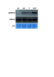
- Figure 3. Induction of Spi-ERK phosphorylation by thermal and UV stresses. Immunoblot performed with ERK and pERK antibodies on protein extracts from coral nubbins incubated for 30 minutes in control (Cont.), thermal stress (T), UV stress (UV) or a combination of thermal and UV stresses (UV + T) conditions. The amido black total protein staining of the western blot membrane is shown as a loading control.
