Explore
 Validate
 Learn
Learn Immunocytochemistry
Antibody data
- Antibody Data
- Antigen structure
- References [0]
- Comments [0]
- Validations
- Immunocytochemistry [2]
Submit
Validation data
Reference
Comment
Report error
- Product number
- MA5-16308-D550 - Provider product page

- Provider
- Invitrogen Antibodies
- Product name
- beta Tubulin Loading Control Monoclonal Antibody (BT7R), DyLight™ 550
- Antibody type
- Monoclonal
- Antigen
- Synthetic peptide
- Description
- Description: The monoclonal antibody TB28-2 recognizes the kappa light chain of human immunoglobulin. Approximately 22-kDa, the Ig kappa chain is comprised of one variable and one constant region. Two kappa chains together with the heavy chain comprise the whole Ig structure. Expression of Ig kappa is typically found on more than half of B lymphocytes.
- Reactivity
- Human, Mouse, Rat, Chicken/Avian, Rabbit
- Host
- Mouse
- Conjugate
- Yellow dye
- Isotype
- IgG
- Antibody clone number
- BT7R
- Vial size
- 50 µL
- Concentration
- 1 mg/mL
- Storage
- 4° C, do not freeze
No comments: Submit comment
Supportive validation
- Submitted by
- Invitrogen Antibodies (provider)
- Main image

- Experimental details
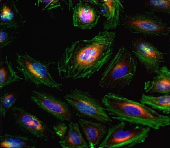
- Immunofluorescent analysis of Beta-Tubulin (red) in HeLa cells. Formalin fixed cells were permeabilized with 0.1% Triton X-100 in TBS for 10 minutes at room temperature and blocked with 1% Blocker BSA (Product # 37525) for 15 minutes at room temperature. Cells were probed with a DyLight 550-conjugated Beta-Tubulin monoclonal antibody (Product # MA5-16308-D550) at a dilution of 1:50 for at least 1 hour at room temperature. F-actin (green) was stained with DyLight 488 Phalloidin (Product # 21833) and nuclei (blue) were stained with Hoechst 33342 dye (Product # 62249). Images were taken on a Thermo Scientific ArrayScan or ToxInsight Instrument at 20X magnification.
- Submitted by
- Invitrogen Antibodies (provider)
- Main image

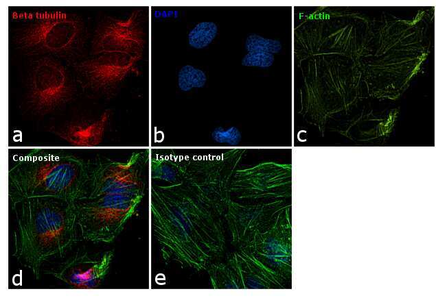
- Experimental details
- Immunofluorescence analysis of beta Tubulin was performed using 70% confluent log phase HeLa cells. The cells were fixed with 4% paraformaldehyde for 10 minutes, permeabilized with 0.1% Triton™ X-100 for 15 minutes, and blocked with 1% BSA for 1 hour at room temperature. The cells were labeled with beta Tubulin Loading Control Monoclonal Antibody (BT7R), DyLight 550 (Product # MA5-16308-D550) at 1:100 dilution in 0.1% BSA and incubated at 4 degree Celsius overnight (Panel a: red). Nuclei (Panel b: blue) were stained with ProLong™ Diamond Antifade Mountant with DAPI (Product # P36962). F-actin (Panel c: green) was stained with Alexa Fluor® 488 Phalloidin (Product # A12379, 1:300). Panel d represents the merged image showing cytoplasmic localization. Panel e represents isotype control to assess background. The images were captured at 60X magnification.