 Explore
 Validate
 Learn
Learn Western blot
Antibody data
- Antibody Data
- Antigen structure
- References [0]
- Comments [0]
- Validations
- Western blot [2]
- Immunocytochemistry [1]
Submit
Validation data
Reference
Comment
Report error
- Product number
- MA5-16308-D680 - Provider product page

- Provider
- Invitrogen Antibodies
- Product name
- beta Tubulin Loading Control Monoclonal Antibody (BT7R), DyLight™ 680
- Antibody type
- Monoclonal
- Antigen
- Synthetic peptide
- Description
- MA5-16308-D680 has successfully been used in immunofluorescence and Western blot applications. DyLight 680 has an excitation/emission of 692/712 nm.
- Reactivity
- Human, Mouse, Rat, Canine, Chicken/Avian, Rabbit
- Host
- Mouse
- Conjugate
- Near infrared dye
- Isotype
- IgG
- Antibody clone number
- BT7R
- Vial size
- 50 µL
- Concentration
- 1 mg/mL
- Storage
- 4° C, do not freeze
No comments: Submit comment
Supportive validation
- Submitted by
- Invitrogen Antibodies (provider)
- Main image
- Experimental details
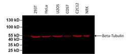
- Western blot analysis of Beta-Tubulin was performed by loading 75 µg of various cell lysates per well onto a 4-20% Tris-HCl polyacrylamide gel. Proteins were transferred to a PVDF membrane and blocked with 5% BSA/TBST for at least 1 hour. The membrane was probed with a DyLight 680-conjugated Beta-Tubulin monoclonal antibody (Product # MA5-16308-D680) at a dilution of 1:1000 for 1 hour at room temperature on a rocking platform and washed in TBS-0.1% Tween-20. Detection was performed using the LI-COR Odyssey.
- Conjugate
- Near infrared dye

- Submitted by
- Invitrogen Antibodies (provider)
- Main image
- Experimental details
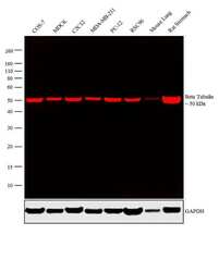
- Western blot analysis was performed on whole cell extracts (30 µg lysate) of COS-7 (Lane 1), MDCK (Lane 2), C2C12 (Lane 3), MDA-MB-231 (Lane 4), PC-12 (Lane 5), RSC96 (Lane 6), tissue extracts of Mouse Lung (Lane 7) and Rat Stomach (Lane 8). The blot was probed with beta Tubulin Loading Control Monoclonal Antibody (BT7R), DyLight 680 (Product # MA5-16308-D680, 1 µg/mL). A 50 kDa band corresponding to beta Tubulin was observed across the cell lines and tissues tested. Known quantity of protein samples were electrophoresed using Novex® NuPAGE® 10% Bis-Tris gel (Product # NP0322BOX), XCell SureLock™ Electrophoresis System (Product # EI0002) and Novex® Sharp Pre-Stained Protein Standard (Product # LC5800). Resolved proteins were then transferred onto a nitrocellulose membrane with iBlot® 2 Dry Blotting System (Product # IB21001). The membrane was probed with the relevant primary antibody following blocking with 5 % skimmed milk. Fluorescent detection was performed using the Odyssey® Fc imaging system (Li-cor Biosciences).
- Conjugate
- Near infrared dye

Supportive validation
- Submitted by
- Invitrogen Antibodies (provider)
- Main image

- Experimental details
- Immunofluorescent analysis of Beta-Tubulin (red) in HeLa cells. Formalin fixed cells were permeabilized with 0.1% Triton X-100 in TBS for 10 minutes at room temperature and blocked with 1% Blocker BSA (Product # 37525) for 15 minutes at room temperature. Cells were probed with a DyLight 680-conjugated Beta-Tubulin monoclonal antibody (Product # MA5-16308-D680) at a dilution of 1:50 for at least 1 hour at room temperature and washed with PBS. F-actin (green) was stained with DyLight 488 Phalloidin (Product # 21833) and nuclei (blue) were stained with Hoechst 33342 dye (Product # 62249). Images were taken on a Thermo Scientific ArrayScan or ToxInsight Instrument at 20X magnification.
- Conjugate
- Near infrared dye
