Explore
 Validate
 Learn
Learn Western blot
Antibody data
- Antibody Data
- Antigen structure
- References [1]
- Comments [0]
- Validations
- Western blot [1]
- Immunocytochemistry [2]
Submit
Validation data
Reference
Comment
Report error
- Product number
- MA5-16308-A647 - Provider product page

- Provider
- Invitrogen Antibodies
- Product name
- beta Tubulin Loading Control Monoclonal Antibody (BT7R), Alexa Fluor™ 647
- Antibody type
- Monoclonal
- Antigen
- Synthetic peptide
- Reactivity
- Human, Mouse, Rat, Chicken/Avian, Rabbit
- Host
- Mouse
- Conjugate
- Red dye
- Isotype
- IgG
- Antibody clone number
- BT7R
- Vial size
- 50 µL
- Concentration
- 1 mg/mL
- Storage
- 4° C, do not freeze
Submitted references Cell Cycle-Dependent Tumor Engraftment and Migration Are Enabled by Aurora-A.
Chu TLH, Connell M, Zhou L, He Z, Won J, Chen H, Rahavi SMR, Mohan P, Nemirovsky O, Fotovati A, Pujana MA, Reid GSD, Nielsen TO, Pante N, Maxwell CA
Molecular cancer research : MCR 2018 Jan;16(1):16-31
Molecular cancer research : MCR 2018 Jan;16(1):16-31
No comments: Submit comment
Supportive validation
- Submitted by
- Invitrogen Antibodies (provider)
- Main image

- Experimental details
- Western blot analysis of beta-Tubulin was performed by loading various cell lysates onto a 4-20% Tris-HCl polyacrylamide gel. Proteins were transferred to a low fluorescence PVDF membrane and blocked with Sea Block blocking buffer for at least 1 hour. The membrane was probed with a AlexaFluor647-conjugated beta-Tubulin monoclonal antibody (Product # MA5-16308-A647) at a dilution of 1:500 for 1 hour at room temperature on a rocking platform and washed in TBS-0.1% Tween-20. Detection was performed using a fluorescence imaging system.
- Conjugate
- Red dye

Supportive validation
- Submitted by
- Invitrogen Antibodies (provider)
- Main image

- Experimental details
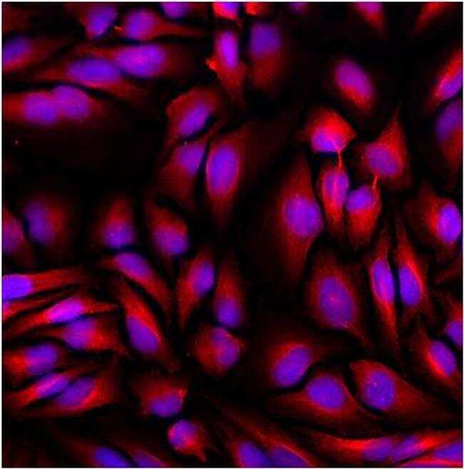
- Immunofluorescent analysis of beta tubulin (red) in HeLa cells. The cells were fixed with 4% paraformaldehyde for 15 minutes, permeabilized with 0.1% Triton X-100 in PBS for 15 minutes and blocked with 3% Blocker BSA (Product # 37525) in PBS for 30 minutes at room temperature. Cells were stained with AlexaFluor 647 conjugated beta-tubulin monoclonal antibody (Product # MA5-16308-A647) at a dilution of 1:50 for 1 hour at room temperature. Nuclei (blue) were stained with Hoechst 33342 dye (Product # 62249). Images were taken on a Thermo Scientific ToxInsight at 20X magnification.
- Conjugate
- Red dye
- Submitted by
- Invitrogen Antibodies (provider)
- Main image
- Experimental details
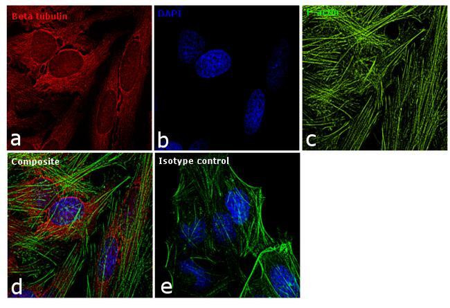
- Immunofluorescence analysis of beta tubulin was performed using 70% confluent log phase HeLa cells. The cells were fixed with 4% paraformaldehyde for 10 minutes, permeabilized with 0.1% Triton™ X-100 for 15 minutes, and blocked with 1% BSA for 1 hour at room temperature. The cells were labeled with beta Tubulin Loading Control Monoclonal Antibody (BT7R), Alexa Fluor 647 (Product # MA5-16308-A647) at 1:25 dilution in 0.1% BSA and incubated at 4 degree Celsius overnight (Panel a: red). Nuclei (Panel b: blue) were stained with SlowFade® Gold Antifade Mountant with DAPI (Product # S36938). F-actin (Panel c: green) was stained with Alexa Fluor® 488 Phalloidin (Product # A12379, 1:300). Panel d represents the merged image showing cytoplasmic localization. Panel e shows the isotype control. The images were captured at 60X magnification.
- Conjugate
- Red dye