Explore
 Validate
 Learn
Learn Western blot
 ELISA
ELISAAntibody data
- Antibody Data
- Antigen structure
- References [73]
- Comments [0]
- Validations
- Western blot [5]
- Immunocytochemistry [5]
- Immunohistochemistry [3]
- Flow cytometry [3]
- Other assay [16]
Submit
Validation data
Reference
Comment
Report error
- Product number
- MA5-16308 - Provider product page

- Provider
- Invitrogen Antibodies
- Product name
- beta Tubulin Loading Control Monoclonal Antibody (BT7R)
- Antibody type
- Monoclonal
- Antigen
- Synthetic peptide
- Reactivity
- Human, Mouse, Rat, Canine, Chicken/Avian, Rabbit
- Host
- Mouse
- Isotype
- IgG
- Antibody clone number
- BT7R
- Vial size
- 100 µg
- Concentration
- 1 mg/mL
- Storage
- -20° C, Avoid Freeze/Thaw Cycles
Submitted references A tubulin binding molecule drives differentiation of acute myeloid leukemia cells.
Oxidative Stress Resistance 1 Gene Therapy Retards Neurodegeneration in the rd1 Mutant Mouse Model of Retinopathy.
Therapeutic Benefit of Galectin-1: Beyond Membrane Repair, a Multifaceted Approach to LGMD2B.
Establishment of an induced pluripotent stem (iPS) cell line (SDUKIi006-A) from a 21-year old male patient diagnosed with atypical autism disorder.
A Cystine-Cysteine Intercellular Shuttle Prevents Ferroptosis in xCT(KO) Pancreatic Ductal Adenocarcinoma Cells.
Overcoming Culture Restriction for SARS-CoV-2 in Human Cells Facilitates the Screening of Compounds Inhibiting Viral Replication.
Adipose Tissue-Derived Mesenchymal Stem Cell Concentrated Conditioned Medium Alters the Expression Pattern of Glutamate Regulatory Proteins and Aquaporin-4 in the Retina after Mild Traumatic Brain Injury.
Hypoxia reduces cell attachment of SARS-CoV-2 spike protein by modulating the expression of ACE2, neuropilin-1, syndecan-1 and cellular heparan sulfate.
Global proteomic analysis of insulin receptor interactors in glomerular podocytes.
Treatment with galectin-1 improves myogenic potential and membrane repair in dysferlin-deficient models.
Hypoxia Promotes Syndecan-3 Expression in the Tumor Microenvironment.
High levels of MESP1 expression in non-small cell lung cancer can facilitate cell proliferation, metastasis and suppresses cell apoptosis.
Derivation of induced pluripotent stem cells (SDUKIi003-A) from a 20-year-old male patient diagnosed with Asperger syndrome.
Royal jelly attenuates metabolic defects in a Drosophila mutant with elevated TORC1 activity.
G(q/11)-dependent regulation of endosomal cAMP generation by parathyroid hormone class B GPCR.
Effect of Melatonin on Tau aggregation and Tau-mediated cell surface morphology.
Generation of human induced pluripotent stem cells (SDUKIi002-A) from a 22-year-old male diagnosed with autism spectrum disorder.
Melatonin Reduces GSK3β-Mediated Tau Phosphorylation, Enhances Nrf2 Nuclear Translocation and Anti-Inflammation.
Precision cut lung slices: a novel versatile tool to examine host-pathogen interaction in the chicken lung.
Synergistic interactions of PlGF and VEGF contribute to blood-retinal barrier breakdown through canonical NFκB activation.
Exploring the Mitochondrial Degradome by the TAILS Proteomics Approach in a Cellular Model of Parkinson's Disease.
Irisin Exerts Inhibitory Effect on Adipogenesis Through Regulation of Wnt Signaling.
HtrA4 Protease Promotes Chemotherapeutic-Dependent Cancer Cell Death.
Collagen XIII-derived ectodomain regulates bone angiogenesis and intracortical remodeling.
Genome-wide effect of pulmonary airway epithelial cell-specific Bmal1 deletion.
GRP78 translocation to the cell surface and O-GlcNAcylation of VE-Cadherin contribute to ER stress-mediated endothelial permeability.
Fetal Gene Therapy Using a Single Injection of Recombinant AAV9 Rescued SMA Phenotype in Mice.
A Genome-wide CRISPR Screen Identifies ZCCHC14 as a Host Factor Required for Hepatitis B Surface Antigen Production.
Sensory innervation in porous endplates by Netrin-1 from osteoclasts mediates PGE2-induced spinal hypersensitivity in mice.
In Preclinical Model of Ovarian Cancer, the SGK1 Inhibitor SI113 Counteracts the Development of Paclitaxel Resistance and Restores Drug Sensitivity.
Genetic Ablation of the Cystine Transporter xCT in PDAC Cells Inhibits mTORC1, Growth, Survival, and Tumor Formation via Nutrient and Oxidative Stresses.
Claudin-19 mediates the effects of NO on the paracellular pathway in thick ascending limbs.
Inhibition of the amino-acid transporter LAT1 demonstrates anti-neoplastic activity in medulloblastoma.
Elevated WBP2 Expression in HER2-positive Breast Cancers Correlates with Sensitivity to Trastuzumab-based Neoadjuvant Therapy: A Retrospective and Multicentric Study.
Downregulation of N-Acetylglucosaminyltransferase GCNT3 by miR-302b-3p Decreases Non-Small Cell Lung Cancer (NSCLC) Cell Proliferation, Migration and Invasion.
Modulation of Autophagy by a Small Molecule Inverse Agonist of ERRα Is Neuroprotective.
Antinociceptive effect of Valeriana fauriei regulates BDNF signaling in an animal model of fibromyalgia.
The glutamine transporter ASCT2 (SLC1A5) promotes tumor growth independently of the amino acid transporter LAT1 (SLC7A5).
Featured Article: Deterioration of visual function mediated by senescence-associated endoplasmic reticulum stress in inflammatory tie2-TNF mice.
Inhibition of PirB Activity by TAT-PEP Improves Mouse Motor Ability and Cognitive Behavior.
Dopamine negatively modulates the NCA ion channels in C. elegans.
The dense-core vesicle maturation protein CCCP-1 binds RAB-2 and membranes through its C-terminal domain.
Cancer progression by reprogrammed BCAA metabolism in myeloid leukaemia.
H(2)S and homocysteine control a novel feedback regulation of cystathionine beta synthase and cystathionine gamma lyase in cardiomyocytes.
A novel autophagy modulator 6-Bio ameliorates SNCA/α-synuclein toxicity.
A START-domain-containing protein is a novel marker of nervous system components of the sea cucumber Holothuria glaberrima.
Gold nanoclusters-assisted delivery of NGF siRNA for effective treatment of pancreatic cancer.
The EARP Complex and Its Interactor EIPR-1 Are Required for Cargo Sorting to Dense-Core Vesicles.
Genetic Disruption of the Multifunctional CD98/LAT1 Complex Demonstrates the Key Role of Essential Amino Acid Transport in the Control of mTORC1 and Tumor Growth.
Prion-like propagation of human brain-derived alpha-synuclein in transgenic mice expressing human wild-type alpha-synuclein.
Early adaptive response of the retina to a pro-diabetogenic diet: Impairment of cone response and gene expression changes in high-fructose fed rats.
The tyrosine phosphatase PTPN14 is a negative regulator of YAP activity.
Opposite effects of a high-fat diet and calorie restriction on ciliary neurotrophic factor signaling in the mouse hypothalamus.
Inositol 1,4,5-trisphosphate receptor regulates replication, differentiation, infectivity and virulence of the parasitic protist Trypanosoma cruzi.
The pseudophosphatase MK-STYX inhibits stress granule assembly independently of Ser149 phosphorylation of G3BP-1.
STAT1 activation by venous malformations mutant Tie2-R849W antagonizes VEGF-A-mediated angiogenic response partly via reduced bFGF production.
Carvedilol treatment after myocardial infarct decreases cardiomyocytic apoptosis in the peri-infarct zone during cardioplegia-induced cardiac arrest.
Oxidative stress, Nrf2 and keratin up-regulation associate with Mallory-Denk body formation in mouse erythropoietic protoporphyria.
MicroRNA-27a regulates cardiomyocytic apoptosis during cardioplegia-induced cardiac arrest by targeting interleukin 10-related pathways.
Indirubin, an acting component of indigo naturalis, inhibits EGFR activation and EGF-induced CDC25B gene expression in epidermal keratinocytes.
DNA methyltransferase 1, cytosine methylation, and cytosine hydroxymethylation in mammalian mitochondria.
Interdependence of platelet-derived growth factor and estrogen-signaling pathways in inducing neonatal rat testicular gonocytes proliferation.
Grape seed extract induces cell cycle arrest and apoptosis in human colon carcinoma cells.
Constitutive overexpression of CDC25A in primary human mammary epithelial cells results in both defective DNA damage response and chromosomal breaks at fragile sites.
Efficient coupling of Sec23-Sec24 to Sec13-Sec31 drives COPII-dependent collagen secretion and is essential for normal craniofacial development.
Growth control of multiple myeloma cells through inhibition of glycogen synthase kinase-3.
Neural differentiation arrest in embryonal carcinoma cells with forced expression of EWS-FLI1.
Endothelial nitric oxide synthase in bicuspid aortic valve disease.
Stimulation of non-transferrin iron uptake by iron deprivation in K562 cells.
Erbin regulates mitogen-activated protein (MAP) kinase activation and MAP kinase-dependent interactions between Merlin and adherens junction protein complexes in Schwann cells.
Mechanical stretch induces podocyte hypertrophy in vitro.
TorsinA negatively controls neurite outgrowth of SH-SY5Y human neuronal cell line.
Nicotine strongly activates dendritic cell-mediated adaptive immunity: potential role for progression of atherosclerotic lesions.
Jackson TR, Vuorinen A, Josa-Culleré L, Madden KS, Conole D, Cogswell TJ, Wilkinson IVL, Kettyle LM, Zhang D, O'Mahony A, Gracias D, McCall L, Westwood R, Terstappen GC, Davies SG, Tate EW, Wynne GM, Vyas P, Russell AJ, Milne TA
iScience 2022 Aug 19;25(8):104787
iScience 2022 Aug 19;25(8):104787
Oxidative Stress Resistance 1 Gene Therapy Retards Neurodegeneration in the rd1 Mutant Mouse Model of Retinopathy.
Sahu B, Leon LM, Zhang W, Puranik N, Periasamy R, Khanna H, Volkert M
Investigative ophthalmology & visual science 2021 Sep 2;62(12):8
Investigative ophthalmology & visual science 2021 Sep 2;62(12):8
Therapeutic Benefit of Galectin-1: Beyond Membrane Repair, a Multifaceted Approach to LGMD2B.
Vallecillo-Zúniga ML, Poulson PD, Luddington JS, Arnold CJ, Rathgeber M, Kartchner BC, Hayes S, Gill H, Valdoz JC, Spallino JL, Garfield S, Dodson EL, Arthur CM, Stowell SR, Van Ry PM
Cells 2021 Nov 17;10(11)
Cells 2021 Nov 17;10(11)
Establishment of an induced pluripotent stem (iPS) cell line (SDUKIi006-A) from a 21-year old male patient diagnosed with atypical autism disorder.
Kamand M, Forsberg SL, Thomassen M, Ilieva M, Meyer M, Svenningsen ÅF, Michel TM
Stem cell research 2021 Mar;51:102185
Stem cell research 2021 Mar;51:102185
A Cystine-Cysteine Intercellular Shuttle Prevents Ferroptosis in xCT(KO) Pancreatic Ductal Adenocarcinoma Cells.
Meira W, Daher B, Parks SK, Cormerais Y, Durivault J, Tambutte E, Pouyssegur J, Vučetić M
Cancers 2021 Mar 21;13(6)
Cancers 2021 Mar 21;13(6)
Overcoming Culture Restriction for SARS-CoV-2 in Human Cells Facilitates the Screening of Compounds Inhibiting Viral Replication.
Ramirez S, Fernandez-Antunez C, Galli A, Underwood A, Pham LV, Ryberg LA, Feng S, Pedersen MS, Mikkelsen LS, Belouzard S, Dubuisson J, Sølund C, Weis N, Gottwein JM, Fahnøe U, Bukh J
Antimicrobial agents and chemotherapy 2021 Jun 17;65(7):e0009721
Antimicrobial agents and chemotherapy 2021 Jun 17;65(7):e0009721
Adipose Tissue-Derived Mesenchymal Stem Cell Concentrated Conditioned Medium Alters the Expression Pattern of Glutamate Regulatory Proteins and Aquaporin-4 in the Retina after Mild Traumatic Brain Injury.
Jha KA, Gentry J, Del Mar NA, Reiner A, Sohl N, Gangaraju R
Journal of neurotrauma 2021 Jun 15;38(12):1702-1716
Journal of neurotrauma 2021 Jun 15;38(12):1702-1716
Hypoxia reduces cell attachment of SARS-CoV-2 spike protein by modulating the expression of ACE2, neuropilin-1, syndecan-1 and cellular heparan sulfate.
Prieto-Fernández E, Egia-Mendikute L, Vila-Vecilla L, Bosch A, Barreira-Manrique A, Lee SY, García-Del Río A, Antoñana-Vildosola A, Jiménez-Lasheras B, Moreno-Cugnon L, Jiménez-Barbero J, Berra E, Ereño-Orbea J, Palazon A
Emerging microbes & infections 2021 Dec;10(1):1065-1076
Emerging microbes & infections 2021 Dec;10(1):1065-1076
Global proteomic analysis of insulin receptor interactors in glomerular podocytes.
Hosawi SB, Humphries JD, Coward RJ, Knight D, Humphries MJ, Lennon R
Wellcome open research 2020;5:202
Wellcome open research 2020;5:202
Treatment with galectin-1 improves myogenic potential and membrane repair in dysferlin-deficient models.
Vallecillo-Zúniga ML, Rathgeber MF, Poulson PD, Hayes S, Luddington JS, Gill HN, Teynor M, Kartchner BC, Valdoz J, Stowell C, Markham AR, Arthur C, Stowell S, Van Ry PM
PloS one 2020;15(9):e0238441
PloS one 2020;15(9):e0238441
Hypoxia Promotes Syndecan-3 Expression in the Tumor Microenvironment.
Prieto-Fernández E, Egia-Mendikute L, Bosch A, García Del Río A, Jimenez-Lasheras B, Antoñana-Vildosola A, Lee SY, Palazon A
Frontiers in immunology 2020;11:586977
Frontiers in immunology 2020;11:586977
High levels of MESP1 expression in non-small cell lung cancer can facilitate cell proliferation, metastasis and suppresses cell apoptosis.
Wang L, Yang C, Li F, Mu D, Ran P, Shen H, Li W, Ma J, Wu J, Yang X, Sheng X, Zhu B, Zheng S
Translational cancer research 2020 Oct;9(10):5956-5968
Translational cancer research 2020 Oct;9(10):5956-5968
Derivation of induced pluripotent stem cells (SDUKIi003-A) from a 20-year-old male patient diagnosed with Asperger syndrome.
Kamand M, Ilieva M, Louise Forsberg S, Thomassen M, Meyer M, Fex Svenningsen Å, Maria Michel T
Stem cell research 2020 Oct;48:101974
Stem cell research 2020 Oct;48:101974
Royal jelly attenuates metabolic defects in a Drosophila mutant with elevated TORC1 activity.
Cheng Y, Cai J, Fu Y, Feng C, Hao Y, Wei Y
Biology open 2020 Nov 6;9(11)
Biology open 2020 Nov 6;9(11)
G(q/11)-dependent regulation of endosomal cAMP generation by parathyroid hormone class B GPCR.
White AD, Jean-Alphonse FG, Fang F, Peña KA, Liu S, König GM, Inoue A, Aslanoglou D, Gellman SH, Kostenis E, Xiao K, Vilardaga JP
Proceedings of the National Academy of Sciences of the United States of America 2020 Mar 31;117(13):7455-7460
Proceedings of the National Academy of Sciences of the United States of America 2020 Mar 31;117(13):7455-7460
Effect of Melatonin on Tau aggregation and Tau-mediated cell surface morphology.
Das R, Balmik AA, Chinnathambi S
International journal of biological macromolecules 2020 Jun 1;152:30-39
International journal of biological macromolecules 2020 Jun 1;152:30-39
Generation of human induced pluripotent stem cells (SDUKIi002-A) from a 22-year-old male diagnosed with autism spectrum disorder.
Kamand M, Ilieva M, Forsberg SL, Thomassen M, Fex Svenningsen Å, Holst B, Meyer M, Michel TM
Stem cell research 2020 Jul;46:101834
Stem cell research 2020 Jul;46:101834
Melatonin Reduces GSK3β-Mediated Tau Phosphorylation, Enhances Nrf2 Nuclear Translocation and Anti-Inflammation.
Das R, Balmik AA, Chinnathambi S
ASN neuro 2020 Jan-Dec;12:1759091420981204
ASN neuro 2020 Jan-Dec;12:1759091420981204
Precision cut lung slices: a novel versatile tool to examine host-pathogen interaction in the chicken lung.
Bryson KJ, Garrido D, Esposito M, McLachlan G, Digard P, Schouler C, Guabiraba R, Trapp S, Vervelde L
Veterinary research 2020 Jan 10;51(1):2
Veterinary research 2020 Jan 10;51(1):2
Synergistic interactions of PlGF and VEGF contribute to blood-retinal barrier breakdown through canonical NFκB activation.
Lennikov A, Mukwaya A, Fan L, Saddala MS, De Falco S, Huang H
Experimental cell research 2020 Dec 15;397(2):112347
Experimental cell research 2020 Dec 15;397(2):112347
Exploring the Mitochondrial Degradome by the TAILS Proteomics Approach in a Cellular Model of Parkinson's Disease.
Lualdi M, Ronci M, Zilocchi M, Corno F, Turilli ES, Sponchiado M, Aceto A, Alberio T, Fasano M
Frontiers in aging neuroscience 2019;11:195
Frontiers in aging neuroscience 2019;11:195
Irisin Exerts Inhibitory Effect on Adipogenesis Through Regulation of Wnt Signaling.
Ma EB, Sahar NE, Jeong M, Huh JY
Frontiers in physiology 2019;10:1085
Frontiers in physiology 2019;10:1085
HtrA4 Protease Promotes Chemotherapeutic-Dependent Cancer Cell Death.
Wenta T, Rychlowski M, Jarzab M, Lipinska B
Cells 2019 Sep 20;8(10)
Cells 2019 Sep 20;8(10)
Collagen XIII-derived ectodomain regulates bone angiogenesis and intracortical remodeling.
Koivunen J, Kemppainen AV, Finnilä MA, Keski-Filppula R, Härönen H, Tu H, Pellikka H, Heikkinen A, Kylmäoja E, Sormunen R, Miinalainen I, Saarakkala S, Izzi V, Pihlajaniemi T
Matrix biology : journal of the International Society for Matrix Biology 2019 Oct;83:6-25
Matrix biology : journal of the International Society for Matrix Biology 2019 Oct;83:6-25
Genome-wide effect of pulmonary airway epithelial cell-specific Bmal1 deletion.
Zhang Z, Hunter L, Wu G, Maidstone R, Mizoro Y, Vonslow R, Fife M, Hopwood T, Begley N, Saer B, Wang P, Cunningham P, Baxter M, Durrington H, Blaikley JF, Hussell T, Rattray M, Hogenesch JB, Gibbs J, Ray DW, Loudon ASI
FASEB journal : official publication of the Federation of American Societies for Experimental Biology 2019 May;33(5):6226-6238
FASEB journal : official publication of the Federation of American Societies for Experimental Biology 2019 May;33(5):6226-6238
GRP78 translocation to the cell surface and O-GlcNAcylation of VE-Cadherin contribute to ER stress-mediated endothelial permeability.
Lenin R, Nagy PG, Jha KA, Gangaraju R
Scientific reports 2019 Jul 25;9(1):10783
Scientific reports 2019 Jul 25;9(1):10783
Fetal Gene Therapy Using a Single Injection of Recombinant AAV9 Rescued SMA Phenotype in Mice.
Rashnonejad A, Amini Chermahini G, Gündüz C, Onay H, Aykut A, Durmaz B, Baka M, Su Q, Gao G, Özkınay F
Molecular therapy : the journal of the American Society of Gene Therapy 2019 Dec 4;27(12):2123-2133
Molecular therapy : the journal of the American Society of Gene Therapy 2019 Dec 4;27(12):2123-2133
A Genome-wide CRISPR Screen Identifies ZCCHC14 as a Host Factor Required for Hepatitis B Surface Antigen Production.
Hyrina A, Jones C, Chen D, Clarkson S, Cochran N, Feucht P, Hoffman G, Lindeman A, Russ C, Sigoillot F, Tsang T, Uehara K, Xie L, Ganem D, Holdorf M
Cell reports 2019 Dec 3;29(10):2970-2978.e6
Cell reports 2019 Dec 3;29(10):2970-2978.e6
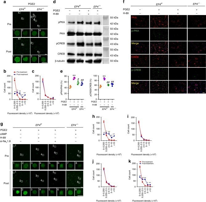
Sensory innervation in porous endplates by Netrin-1 from osteoclasts mediates PGE2-induced spinal hypersensitivity in mice.
Ni S, Ling Z, Wang X, Cao Y, Wu T, Deng R, Crane JL, Skolasky R, Demehri S, Zhen G, Jain A, Wu P, Pan D, Hu B, Lyu X, Li Y, Chen H, Qi H, Guan Y, Dong X, Wan M, Zou X, Lu H, Hu J, Cao X
Nature communications 2019 Dec 10;10(1):5643
Nature communications 2019 Dec 10;10(1):5643
In Preclinical Model of Ovarian Cancer, the SGK1 Inhibitor SI113 Counteracts the Development of Paclitaxel Resistance and Restores Drug Sensitivity.
D'Antona L, Dattilo V, Catalogna G, Scumaci D, Fiumara CV, Musumeci F, Perrotti G, Schenone S, Tallerico R, Spoleti CB, Costa N, Iuliano R, Cuda G, Amato R, Perrotti N
Translational oncology 2019 Aug;12(8):1045-1055
Translational oncology 2019 Aug;12(8):1045-1055
Genetic Ablation of the Cystine Transporter xCT in PDAC Cells Inhibits mTORC1, Growth, Survival, and Tumor Formation via Nutrient and Oxidative Stresses.
Daher B, Parks SK, Durivault J, Cormerais Y, Baidarjad H, Tambutte E, Pouysségur J, Vučetić M
Cancer research 2019 Aug 1;79(15):3877-3890
Cancer research 2019 Aug 1;79(15):3877-3890
Claudin-19 mediates the effects of NO on the paracellular pathway in thick ascending limbs.
Monzon CM, Garvin JL
American journal of physiology. Renal physiology 2019 Aug 1;317(2):F411-F418
American journal of physiology. Renal physiology 2019 Aug 1;317(2):F411-F418
Inhibition of the amino-acid transporter LAT1 demonstrates anti-neoplastic activity in medulloblastoma.
Cormerais Y, Pagnuzzi-Boncompagni M, Schrötter S, Giuliano S, Tambutté E, Endou H, Wempe MF, Pagès G, Pouysségur J, Picco V
Journal of cellular and molecular medicine 2019 Apr;23(4):2711-2718
Journal of cellular and molecular medicine 2019 Apr;23(4):2711-2718
Elevated WBP2 Expression in HER2-positive Breast Cancers Correlates with Sensitivity to Trastuzumab-based Neoadjuvant Therapy: A Retrospective and Multicentric Study.
Kang SA, Guan JS, Tan HJ, Chu T, Thike AA, Bernadó C, Arribas J, Wong CY, Tan PH, Gudi M, Putti TC, Sohn J, Lim SH, Lee SC, Lim YP
Clinical cancer research : an official journal of the American Association for Cancer Research 2019 Apr 15;25(8):2588-2600
Clinical cancer research : an official journal of the American Association for Cancer Research 2019 Apr 15;25(8):2588-2600
Downregulation of N-Acetylglucosaminyltransferase GCNT3 by miR-302b-3p Decreases Non-Small Cell Lung Cancer (NSCLC) Cell Proliferation, Migration and Invasion.
Li Q, Ran P, Zhang X, Guo X, Yuan Y, Dong T, Zhu B, Zheng S, Xiao C
Cellular physiology and biochemistry : international journal of experimental cellular physiology, biochemistry, and pharmacology 2018;50(3):987-1004
Cellular physiology and biochemistry : international journal of experimental cellular physiology, biochemistry, and pharmacology 2018;50(3):987-1004
Modulation of Autophagy by a Small Molecule Inverse Agonist of ERRα Is Neuroprotective.
Suresh SN, Chavalmane AK, Pillai M, Ammanathan V, Vidyadhara DJ, Yarreiphang H, Rai S, Paul A, Clement JP, Alladi PA, Manjithaya R
Frontiers in molecular neuroscience 2018;11:109
Frontiers in molecular neuroscience 2018;11:109
Antinociceptive effect of Valeriana fauriei regulates BDNF signaling in an animal model of fibromyalgia.
Lee H, Im J, Won H, Kim JY, Kim HK, Kwon JT, Kim YO, Lee S, Cho IH, Lee SW, Kim HJ
International journal of molecular medicine 2018 Jan;41(1):485-492
International journal of molecular medicine 2018 Jan;41(1):485-492
The glutamine transporter ASCT2 (SLC1A5) promotes tumor growth independently of the amino acid transporter LAT1 (SLC7A5).
Cormerais Y, Massard PA, Vucetic M, Giuliano S, Tambutté E, Durivault J, Vial V, Endou H, Wempe MF, Parks SK, Pouyssegur J
The Journal of biological chemistry 2018 Feb 23;293(8):2877-2887
The Journal of biological chemistry 2018 Feb 23;293(8):2877-2887
Featured Article: Deterioration of visual function mediated by senescence-associated endoplasmic reticulum stress in inflammatory tie2-TNF mice.
Lenin R, Nagy PG, Gentry J, Gangaraju R
Experimental biology and medicine (Maywood, N.J.) 2018 Aug;243(12):976-984
Experimental biology and medicine (Maywood, N.J.) 2018 Aug;243(12):976-984
Inhibition of PirB Activity by TAT-PEP Improves Mouse Motor Ability and Cognitive Behavior.
Mi YJ, Chen H, Guo N, Sun MY, Zhao ZH, Gao XC, Wang XL, Zhang RS, Zhou JB, Gou XC
Frontiers in aging neuroscience 2017;9:199
Frontiers in aging neuroscience 2017;9:199
Dopamine negatively modulates the NCA ion channels in C. elegans.
Topalidou I, Cooper K, Pereira L, Ailion M
PLoS genetics 2017 Oct;13(10):e1007032
PLoS genetics 2017 Oct;13(10):e1007032
The dense-core vesicle maturation protein CCCP-1 binds RAB-2 and membranes through its C-terminal domain.
Cattin-Ortolá J, Topalidou I, Dosey A, Merz AJ, Ailion M
Traffic (Copenhagen, Denmark) 2017 Nov;18(11):720-732
Traffic (Copenhagen, Denmark) 2017 Nov;18(11):720-732
Cancer progression by reprogrammed BCAA metabolism in myeloid leukaemia.
Hattori A, Tsunoda M, Konuma T, Kobayashi M, Nagy T, Glushka J, Tayyari F, McSkimming D, Kannan N, Tojo A, Edison AS, Ito T
Nature 2017 May 25;545(7655):500-504
Nature 2017 May 25;545(7655):500-504
H(2)S and homocysteine control a novel feedback regulation of cystathionine beta synthase and cystathionine gamma lyase in cardiomyocytes.
Nandi SS, Mishra PK
Scientific reports 2017 Jun 16;7(1):3639
Scientific reports 2017 Jun 16;7(1):3639
A novel autophagy modulator 6-Bio ameliorates SNCA/α-synuclein toxicity.
Suresh SN, Chavalmane AK, Dj V, Yarreiphang H, Rai S, Paul A, Clement JP, Alladi PA, Manjithaya R
Autophagy 2017 Jul 3;13(7):1221-1234
Autophagy 2017 Jul 3;13(7):1221-1234
A START-domain-containing protein is a novel marker of nervous system components of the sea cucumber Holothuria glaberrima.
Rosado-Olivieri EA, Ramos-Ortiz GA, Hernández-Pasos J, Díaz-Balzac CA, Vázquez-Rosa E, Valentín-Tirado G, Vega IE, García-Arrarás JE
Comparative biochemistry and physiology. Part B, Biochemistry & molecular biology 2017 Dec;214:57-65
Comparative biochemistry and physiology. Part B, Biochemistry & molecular biology 2017 Dec;214:57-65
Gold nanoclusters-assisted delivery of NGF siRNA for effective treatment of pancreatic cancer.
Lei Y, Tang L, Xie Y, Xianyu Y, Zhang L, Wang P, Hamada Y, Jiang K, Zheng W, Jiang X
Nature communications 2017 Apr 25;8:15130
Nature communications 2017 Apr 25;8:15130
The EARP Complex and Its Interactor EIPR-1 Are Required for Cargo Sorting to Dense-Core Vesicles.
Topalidou I, Cattin-Ortolá J, Pappas AL, Cooper K, Merrihew GE, MacCoss MJ, Ailion M
PLoS genetics 2016 May;12(5):e1006074
PLoS genetics 2016 May;12(5):e1006074
Genetic Disruption of the Multifunctional CD98/LAT1 Complex Demonstrates the Key Role of Essential Amino Acid Transport in the Control of mTORC1 and Tumor Growth.
Cormerais Y, Giuliano S, LeFloch R, Front B, Durivault J, Tambutté E, Massard PA, de la Ballina LR, Endou H, Wempe MF, Palacin M, Parks SK, Pouyssegur J
Cancer research 2016 Aug 1;76(15):4481-92
Cancer research 2016 Aug 1;76(15):4481-92
Prion-like propagation of human brain-derived alpha-synuclein in transgenic mice expressing human wild-type alpha-synuclein.
Bernis ME, Babila JT, Breid S, Wüsten KA, Wüllner U, Tamgüney G
Acta neuropathologica communications 2015 Nov 26;3:75
Acta neuropathologica communications 2015 Nov 26;3:75
Early adaptive response of the retina to a pro-diabetogenic diet: Impairment of cone response and gene expression changes in high-fructose fed rats.
Thierry M, Pasquis B, Buteau B, Fourgeux C, Dembele D, Leclere L, Gambert-Nicot S, Acar N, Bron AM, Creuzot-Garcher CP, Bretillon L
Experimental eye research 2015 Jun;135:37-46
Experimental eye research 2015 Jun;135:37-46
The tyrosine phosphatase PTPN14 is a negative regulator of YAP activity.
Michaloglou C, Lehmann W, Martin T, Delaunay C, Hueber A, Barys L, Niu H, Billy E, Wartmann M, Ito M, Wilson CJ, Digan ME, Bauer A, Voshol H, Christofori G, Sellers WR, Hofmann F, Schmelzle T
PloS one 2013;8(4):e61916
PloS one 2013;8(4):e61916
Opposite effects of a high-fat diet and calorie restriction on ciliary neurotrophic factor signaling in the mouse hypothalamus.
Severi I, Perugini J, Mondini E, Smorlesi A, Frontini A, Cinti S, Giordano A
Frontiers in neuroscience 2013;7:263
Frontiers in neuroscience 2013;7:263
Inositol 1,4,5-trisphosphate receptor regulates replication, differentiation, infectivity and virulence of the parasitic protist Trypanosoma cruzi.
Hashimoto M, Enomoto M, Morales J, Kurebayashi N, Sakurai T, Hashimoto T, Nara T, Mikoshiba K
Molecular microbiology 2013 Mar;87(6):1133-50
Molecular microbiology 2013 Mar;87(6):1133-50
The pseudophosphatase MK-STYX inhibits stress granule assembly independently of Ser149 phosphorylation of G3BP-1.
Barr JE, Munyikwa MR, Frazier EA, Hinton SD
The FEBS journal 2013 Jan;280(1):273-84
The FEBS journal 2013 Jan;280(1):273-84
STAT1 activation by venous malformations mutant Tie2-R849W antagonizes VEGF-A-mediated angiogenic response partly via reduced bFGF production.
Huang YH, Wu MP, Pan SC, Su WC, Chen YW, Wu LW
Angiogenesis 2013 Jan;16(1):207-22
Angiogenesis 2013 Jan;16(1):207-22
Carvedilol treatment after myocardial infarct decreases cardiomyocytic apoptosis in the peri-infarct zone during cardioplegia-induced cardiac arrest.
Yeh CH, Chen TP, Wang YC, Lin YM, Fang SW
Shock (Augusta, Ga.) 2013 Apr;39(4):343-52
Shock (Augusta, Ga.) 2013 Apr;39(4):343-52
Oxidative stress, Nrf2 and keratin up-regulation associate with Mallory-Denk body formation in mouse erythropoietic protoporphyria.
Singla A, Moons DS, Snider NT, Wagenmaker ER, Jayasundera VB, Omary MB
Hepatology (Baltimore, Md.) 2012 Jul;56(1):322-31
Hepatology (Baltimore, Md.) 2012 Jul;56(1):322-31
MicroRNA-27a regulates cardiomyocytic apoptosis during cardioplegia-induced cardiac arrest by targeting interleukin 10-related pathways.
Yeh CH, Chen TP, Wang YC, Lin YM, Fang SW
Shock (Augusta, Ga.) 2012 Dec;38(6):607-14
Shock (Augusta, Ga.) 2012 Dec;38(6):607-14
Indirubin, an acting component of indigo naturalis, inhibits EGFR activation and EGF-induced CDC25B gene expression in epidermal keratinocytes.
Hsieh WL, Lin YK, Tsai CN, Wang TM, Chen TY, Pang JH
Journal of dermatological science 2012 Aug;67(2):140-6
Journal of dermatological science 2012 Aug;67(2):140-6
DNA methyltransferase 1, cytosine methylation, and cytosine hydroxymethylation in mammalian mitochondria.
Shock LS, Thakkar PV, Peterson EJ, Moran RG, Taylor SM
Proceedings of the National Academy of Sciences of the United States of America 2011 Mar 1;108(9):3630-5
Proceedings of the National Academy of Sciences of the United States of America 2011 Mar 1;108(9):3630-5
Interdependence of platelet-derived growth factor and estrogen-signaling pathways in inducing neonatal rat testicular gonocytes proliferation.
Thuillier R, Mazer M, Manku G, Boisvert A, Wang Y, Culty M
Biology of reproduction 2010 May;82(5):825-36
Biology of reproduction 2010 May;82(5):825-36
Grape seed extract induces cell cycle arrest and apoptosis in human colon carcinoma cells.
Kaur M, Mandair R, Agarwal R, Agarwal C
Nutrition and cancer 2008;60 Suppl 1(Suppl 1):2-11
Nutrition and cancer 2008;60 Suppl 1(Suppl 1):2-11
Constitutive overexpression of CDC25A in primary human mammary epithelial cells results in both defective DNA damage response and chromosomal breaks at fragile sites.
Cangi MG, Piccinin S, Pecciarini L, Talarico A, Dal Cin E, Grassi S, Grizzo A, Maestro R, Doglioni C
International journal of cancer 2008 Sep 15;123(6):1466-71
International journal of cancer 2008 Sep 15;123(6):1466-71
Efficient coupling of Sec23-Sec24 to Sec13-Sec31 drives COPII-dependent collagen secretion and is essential for normal craniofacial development.
Townley AK, Feng Y, Schmidt K, Carter DA, Porter R, Verkade P, Stephens DJ
Journal of cell science 2008 Sep 15;121(Pt 18):3025-34
Journal of cell science 2008 Sep 15;121(Pt 18):3025-34
Growth control of multiple myeloma cells through inhibition of glycogen synthase kinase-3.
Zhou Y, Uddin S, Zimmerman T, Kang JA, Ulaszek J, Wickrema A
Leukemia & lymphoma 2008 Oct;49(10):1945-53
Leukemia & lymphoma 2008 Oct;49(10):1945-53
Neural differentiation arrest in embryonal carcinoma cells with forced expression of EWS-FLI1.
Yang Y, Zhang L, Wei Y, Wang H, Fukuma M, Xu H, Xiong W, Zheng J
Journal of neuro-oncology 2008 Nov;90(2):141-50
Journal of neuro-oncology 2008 Nov;90(2):141-50
Endothelial nitric oxide synthase in bicuspid aortic valve disease.
Aicher D, Urbich C, Zeiher A, Dimmeler S, Schäfers HJ
The Annals of thoracic surgery 2007 Apr;83(4):1290-4
The Annals of thoracic surgery 2007 Apr;83(4):1290-4
Stimulation of non-transferrin iron uptake by iron deprivation in K562 cells.
Kovar J, Neubauerova J, Cimburova M, Truksa J, Balusikova K, Horak J
Blood cells, molecules & diseases 2006 Sep-Oct;37(2):95-9
Blood cells, molecules & diseases 2006 Sep-Oct;37(2):95-9
Erbin regulates mitogen-activated protein (MAP) kinase activation and MAP kinase-dependent interactions between Merlin and adherens junction protein complexes in Schwann cells.
Rangwala R, Banine F, Borg JP, Sherman LS
The Journal of biological chemistry 2005 Mar 25;280(12):11790-7
The Journal of biological chemistry 2005 Mar 25;280(12):11790-7
Mechanical stretch induces podocyte hypertrophy in vitro.
Petermann AT, Pippin J, Durvasula R, Pichler R, Hiromura K, Monkawa T, Couser WG, Shankland SJ
Kidney international 2005 Jan;67(1):157-66
Kidney international 2005 Jan;67(1):157-66
TorsinA negatively controls neurite outgrowth of SH-SY5Y human neuronal cell line.
Ferrari-Toninelli G, Paccioretti S, Francisconi S, Uberti D, Memo M
Brain research 2004 Jun 25;1012(1-2):75-81
Brain research 2004 Jun 25;1012(1-2):75-81
Nicotine strongly activates dendritic cell-mediated adaptive immunity: potential role for progression of atherosclerotic lesions.
Aicher A, Heeschen C, Mohaupt M, Cooke JP, Zeiher AM, Dimmeler S
Circulation 2003 Feb 4;107(4):604-11
Circulation 2003 Feb 4;107(4):604-11
No comments: Submit comment
Supportive validation
- Submitted by
- Invitrogen Antibodies (provider)
- Main image

- Experimental details
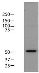
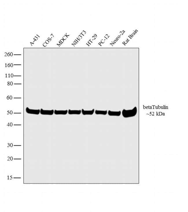
- Western blot analysis was performed on whole cell extracts (30µg lysate) of A-431 (Lane 1), COS-7 (Lane 2), MDCK (Lane 3), NIH/3T3 (Lane 4), HT-29 (Lane 5), PC-12 (Lane 6), Neuro-2a (Lane 7) and tissue extract of Rat Brain (Lane 8). The blot was probed with Anti- beta Tubulin Mouse Monoclonal Antibody (Product # MA5-16308-1MG, 1:2000 dilution) and detected by chemiluminescence using Goat anti-Mouse IgG (H+L) Superclonal™ Secondary Antibody, HRP conjugate (Product # A28177, 0.25µg/mL, 1:4000 dilution). A 52 kDa band corresponding to beta Tubulin was observed across the cell lines and tissue tested. Known quantity of protein samples were electrophoresed using Novex® NuPAGE® 4-12 % Bis-Tris gel (Product # NP0322BOX), XCell SureLock™ Electrophoresis System (Product # EI0002) and Novex® Sharp Pre-Stained Protein Standard (Product # LC5800). Resolved proteins were then transferred onto a nitrocellulose membrane with iBlot® 2 Dry Blotting System (Product # IB21001). The membrane was probed with the relevant primary and secondary Antibody following blocking with 5 % skimmed milk. Chemiluminescent detection was performed using Pierce™ ECL Western Blotting Substrate (Product # 32106).

- Submitted by
- Invitrogen Antibodies (provider)
- Main image
- Experimental details
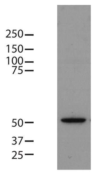
- Western blot analysis of beta-tubulin was performed by loading 8 µg of mouse embryonic stem cell lysate onto an SDS-PAGE gel. Proteins were transferred to a PVDF membrane and blocked with 5% milk in TBST for one hour at room temperature. The membrane was probed with a beta-tubulin loading control monoclonal antibody (Product # MA5-16308) at a dilution of 1:3000 for two hours at room temperature, washed in TBST, and probed with an HRP-conjugated anti-mouse IgG secondary antibody at a dilution of 1:25,000 for one hour at room temperature. Detection was performed using a chemiluminescent substrate. Data courtesy of the Innovators Program.
- Submitted by
- Invitrogen Antibodies (provider)
- Main image

- Experimental details
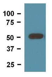
- Western blot analysis of beta-Tubulin was performed by loading 20 µg of THP-1 whole cell lysate per well onto a SDS-PAGE gel. Proteins were transferred to a membrane and blocked with 5% non-fat dry milk in TBST. The membrane was probed with a beta-Tubulin loading control monoclonal antibody (Product # MA5-16308) at a dilution of 1:5000 followed by a HRP-conjugated anti-mouse IgG secondary antibody. Chemiluminescent detection was performed using ECL substrate. Data courtesy of the Innovators Program.

- Submitted by
- Invitrogen Antibodies (provider)
- Main image
- Experimental details
- Western blot of mouse brain tissue lysate loaded at 10 µg/lane and detected with 0.25 µg/mL (Product # MA5-16308).

- Submitted by
- Invitrogen Antibodies (provider)
- Main image

- Experimental details
- Western blot analysis was performed on whole cell extracts (30µg lysate) of A-431 (Lane 1), COS-7 (Lane 2), MDCK (Lane 3), NIH/3T3 (Lane 4), HT-29 (Lane 5), PC-12 (Lane 6), Neuro-2a (Lane 7) and tissue extract of Rat Brain (Lane 8). The blot was probed with Anti- beta Tubulin Mouse Monoclonal Antibody (Product # MA5-16308-1MG, 1:2000 dilution) and detected by chemiluminescence using Goat anti-Mouse IgG (H+L) Superclonal™ Secondary Antibody, HRP conjugate (Product # A28177, 0.25µg/mL, 1:4000 dilution). A 52 kDa band corresponding to beta Tubulin was observed across the cell lines and tissue tested. Known quantity of protein samples were electrophoresed using Novex® NuPAGE® 4-12 % Bis-Tris gel (Product # NP0322BOX), XCell SureLock™ Electrophoresis System (Product # EI0002) and Novex® Sharp Pre-Stained Protein Standard (Product # LC5800). Resolved proteins were then transferred onto a nitrocellulose membrane with iBlot® 2 Dry Blotting System (Product # IB21001). The membrane was probed with the relevant primary and secondary Antibody following blocking with 5 % skimmed milk. Chemiluminescent detection was performed using Pierce™ ECL Western Blotting Substrate (Product # 32106).
Supportive validation
- Submitted by
- Invitrogen Antibodies (provider)
- Main image

- Experimental details
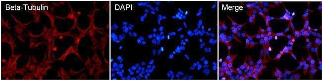
- Immunofluorescent analysis of Beta-Tubulin (red) in HEK293T cells. Cells fixed in 4% formaldehyde were permeabilized and blocked with 1X PBS containing 5% BSA and 0.3% Triton X-100 for 1 hour at room temperature. Cells were probed with a Beta-Tubulin monoclonal antibody (Product # MA5-16308) at a dilution of 1:100 overnight at 4°C in 1X PBS containing 1% BSA and 0.3% Triton X-100, washed with 1X PBS, and incubated with a fluorophore-conjugated goat anti-mouse IgG secondary antibody at a dilution of 1:200 for 1 hour at room temperature. Nuclei (blue) were stained with DAPI. Images were taken on a Leica DM1000 microscope at 40X magnification. Data courtesy of the Innovators Program.
- Submitted by
- Invitrogen Antibodies (provider)
- Main image
- Experimental details
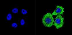
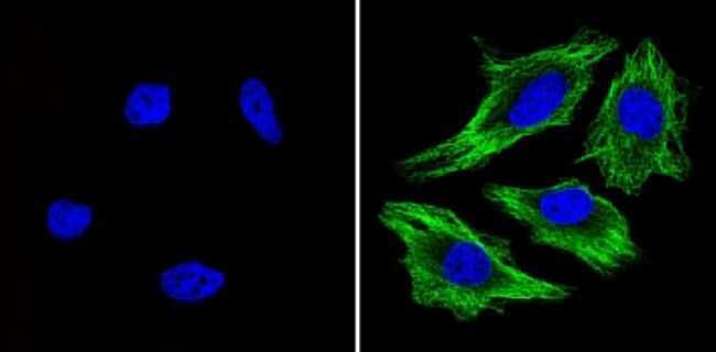
- Immunofluorescent analysis of Beta-Tubulin (green) showing staining in the in the cytoskeleton of A431 cells (right) compared to a negative control without primary antibody (left). Formalin-fixed cells were permeabilized with 0.1% Triton X-100 in TBS for 5-10 minutes and blocked with 3% BSA-PBS for 30 minutes at room temperature. Cells were probed with a Beta-Tubulin loading control antibody (Product # MA5-16308) in 3% BSA-PBS at a dilution of 1:150 and incubated overnight at 4ºC in a humidified chamber. Cells were washed with PBST and incubated with a DyLight-conjugated secondary antibody in PBS at room temperature in the dark. Nuclei (blue) were stained with Hoechst or DAPI. Images were taken at a magnification of 60x.

- Submitted by
- Invitrogen Antibodies (provider)
- Main image

- Experimental details
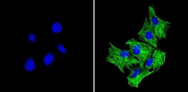
- Immunofluorescent analysis of Beta-Tubulin (green) showing staining in the in the cytoskeleton of C2C12 cells (right) compared to a negative control without primary antibody (left). Formalin-fixed cells were permeabilized with 0.1% Triton X-100 in TBS for 5-10 minutes and blocked with 3% BSA-PBS for 30 minutes at room temperature. Cells were probed with a Beta-Tubulin loading control antibody (Product # MA5-16308) in 3% BSA-PBS at a dilution of 1:150 and incubated overnight at 4ºC in a humidified chamber. Cells were washed with PBST and incubated with a DyLight-conjugated secondary antibody in PBS at room temperature in the dark. Nuclei (blue) were stained with Hoechst or DAPI. Images were taken at a magnification of 60x.
- Submitted by
- Invitrogen Antibodies (provider)
- Main image

- Experimental details
- Immunofluorescent analysis of Beta-Tubulin (green) showing staining in the in the cytoskeleton of Hela cells (right) compared to a negative control without primary antibody (left). Formalin-fixed cells were permeabilized with 0.1% Triton X-100 in TBS for 5-10 minutes and blocked with 3% BSA-PBS for 30 minutes at room temperature. Cells were probed with a Beta-Tubulin loading control antibody (Product # MA5-16308) in 3% BSA-PBS at a dilution of 1:150 and incubated overnight at 4ºC in a humidified chamber. Cells were washed with PBST and incubated with a DyLight-conjugated secondary antibody in PBS at room temperature in the dark. Nuclei (blue) were stained with Hoechst or DAPI. Images were taken at a magnification of 60x.
- Submitted by
- Invitrogen Antibodies (provider)
- Main image
- Experimental details
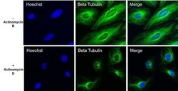
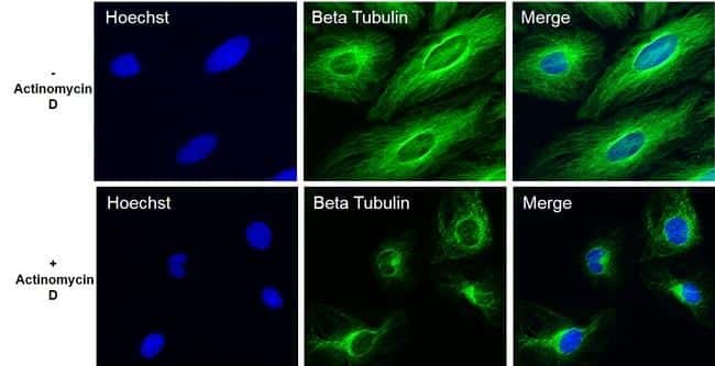
- Immunofluorescent analysis of Beta- Tubulin (green) in HeLa cells untreated or treated with 1uM Actinomycin D for 19 hours. The cells were fixed with 4% Paraformaldehyde for 15 minutes, permeabilized with 0.1% Triton X-100 for 15 minutes, and blocked with 3% BSA for 30 minutes at room temperature. Cells were stained with a Beta-Tubulin mouse monoclonal antibody (Product # MA5-16308) at a concentration of 2.5 µg/mL in blocking buffer for 1 hour at room temperature, and then incubated with a Goat anti-Mouse IgG (H+L) Secondary Antibody, Alexa Fluor Plus 488 conjugate (Product # A32723) at a dilution of 1:500 for at least 30 minutes at a room temperature in the dark (green). Nuclei (blue) were stained with Hoechst 33342 (Product # 62249). Images were taken on a Thermo Scientific ToxInsight Instrument at 20X magnification.
Supportive validation
- Submitted by
- Invitrogen Antibodies (provider)
- Main image
- Experimental details
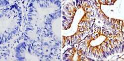
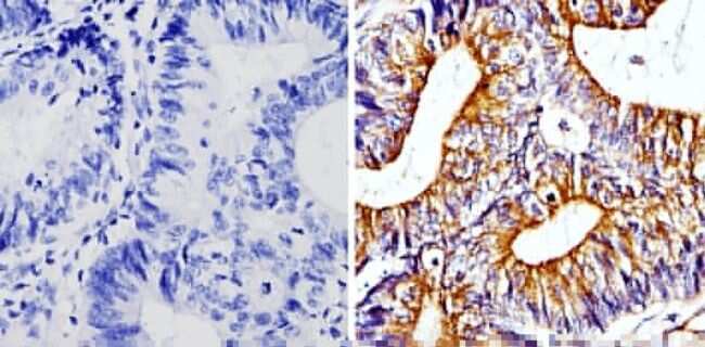
- Immunohistochemistry analysis of Beta-Tubulin showing staining in the cytoskeleton of paraffin-embedded human colon carcinoma (right) compared with a negative control without primary antibody (left). To expose target proteins, antigen retrieval was performed using 10mM sodium citrate (pH 6.0), microwaved for 8-15 min. Following antigen retrieval, tissues were blocked in 3% H2O2-methanol for 15 min at room temperature, washed with ddH2O and PBS, and then probed with a Beta-Tubulin loading control antibody (Product # MA5-16308) diluted in 3% BSA-PBS at a dilution of 1:100 overnight at 4°C in a humidified chamber. Tissues were washed extensively in PBST and detection was performed using an HRP-conjugated secondary antibody followed by colorimetric detection using a DAB kit. Tissues were counterstained with hematoxylin and dehydrated with ethanol and xylene to prep for mounting.
- Submitted by
- Invitrogen Antibodies (provider)
- Main image
- Experimental details
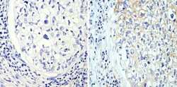
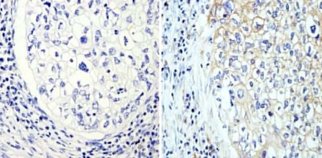
- Immunohistochemistry analysis of Beta-Tubulin showing staining in the cytoskeleton of paraffin-embedded human lung adenocarcinoma (right) compared with a negative control without primary antibody (left). To expose target proteins, antigen retrieval was performed using 10mM sodium citrate (pH 6.0), microwaved for 8-15 min. Following antigen retrieval, tissues were blocked in 3% H2O2-methanol for 15 min at room temperature, washed with ddH2O and PBS, and then probed with a Beta-Tubulin loading control antibody (Product # MA5-16308) diluted in 3% BSA-PBS at a dilution of 1:100 overnight at 4°C in a humidified chamber. Tissues were washed extensively in PBST and detection was performed using an HRP-conjugated secondary antibody followed by colorimetric detection using a DAB kit. Tissues were counterstained with hematoxylin and dehydrated with ethanol and xylene to prep for mounting.
- Submitted by
- Invitrogen Antibodies (provider)
- Main image

- Experimental details
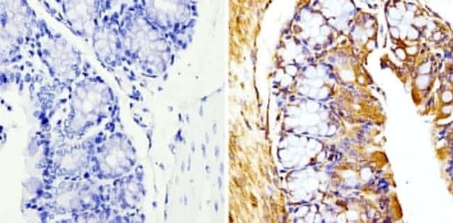
- Immunohistochemistry analysis of Beta-Tubulin showing staining in the cytoskeleton of paraffin-embedded mouse colon tissue (right) compared with a negative control without primary antibody (left). To expose target proteins, antigen retrieval was performed using 10mM sodium citrate (pH 6.0), microwaved for 8-15 min. Following antigen retrieval, tissues were blocked in 3% H2O2-methanol for 15 min at room temperature, washed with ddH2O and PBS, and then probed with a Beta-Tubulin loading control antibody (Product # MA5-16308) diluted in 3% BSA-PBS at a dilution of 1:20 overnight at 4°C in a humidified chamber. Tissues were washed extensively in PBST and detection was performed using an HRP-conjugated secondary antibody followed by colorimetric detection using a DAB kit. Tissues were counterstained with hematoxylin and dehydrated with ethanol and xylene to prep for mounting.
Supportive validation
- Submitted by
- Invitrogen Antibodies (provider)
- Main image
- Experimental details
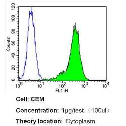
- Flow cytometry analysis of Beta Tubulin in CEM cells (green) compared to an isotype control (blue). Cells were harvested, adjusted to a concentration of 1-5x10^6 cells/mL, fixed with 2% paraformaldehyde and washed with PBS. Cells were blocked with a 2% solution of BSA-PBS for 30 min at room temperature and incubated with a Beta Tubulin loading control antibody (Product # MA5-16308) at a dilution of 1 µg/test for 40 min at room temperature. Cells were then incubated for 40 min at room temperature in the dark using a Dylight 488-conjugated secondary antibody and re-suspended in PBS for FACS analysis.

- Submitted by
- Invitrogen Antibodies (provider)
- Main image
- Experimental details
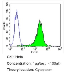
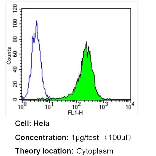
- Flow cytometry analysis of Beta Tubulin in Hela cells (green) compared to an isotype control (blue). Cells were harvested, adjusted to a concentration of 1-5x10^6 cells/mL, fixed with 2% paraformaldehyde and washed with PBS. Cells were blocked with a 2% solution of BSA-PBS for 30 min at room temperature and incubated with a Beta Tubulin loading control antibody (Product # MA5-16308) at a dilution of 1 µg/test for 40 min at room temperature. Cells were then incubated for 40 min at room temperature in the dark using a Dylight 488-conjugated secondary antibody and re-suspended in PBS for FACS analysis.
- Submitted by
- Invitrogen Antibodies (provider)
- Main image
- Experimental details
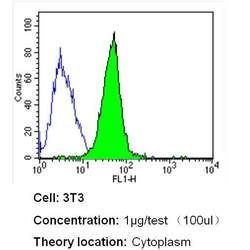
- Flow cytometry analysis of Beta Tubulin in NIH-3T3 cells (green) compared to an isotype control (blue). Cells were harvested, adjusted to a concentration of 1-5x10^6 cells/mL, fixed with 2% paraformaldehyde and washed with PBS. Cells were blocked with a 2% solution of BSA-PBS for 30 min at room temperature and incubated with a Beta Tubulin loading control antibody (Product # MA5-16308) at a dilution of 1 µg/test for 40 min at room temperature. Cells were then incubated for 40 min at room temperature in the dark using a Dylight 488-conjugated secondary antibody and re-suspended in PBS for FACS analysis.

Supportive validation
- Submitted by
- Invitrogen Antibodies (provider)
- Main image

- Experimental details
- NULL
- Submitted by
- Invitrogen Antibodies (provider)
- Main image
- Experimental details
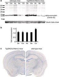
- Fig. 3 Biochemical and histological analysis of alpha-synuclein expression in wild-type and Tg(SNCA)1Nbm/J mice. Biochemical analysis with an antibody against alpha-synuclein (clone 42) that equally reacts with human as well as mouse alpha-synuclein shows that human alpha-synuclein is only very moderately overexpressed in Tg(SNCA)1Nbm/J mice in comparison to alpha-synuclein expression in wild-type mice. Equal amounts of total protein (5 mug) were loaded onto each lane of the SDS-polyacrylamide gel and show expression of alpha-synuclein in the striatum (Str), cortex (Ctx), hippocampus (Hc), brainstem (Bs), and cerebellum (Cb). Molecular sizes are shown in kilodalton a . The signal for alpha-synuclein was quantified by densitometry from western blots and normalized against tubulin and is shown as fold overexpression of alpha-synuclein in Tg(SNCA)1Nbm/J mice (n = 3) versus wild-type mice (n = 3) b . Immunohistochemistry of mouse brain sections with the same antibody (clone 42) against alpha-synuclein reveals comparable localization of alpha-synuclein in Tg(SNCA)1Nbm/J and wild-type mice c

- Submitted by
- Invitrogen Antibodies (provider)
- Main image

- Experimental details
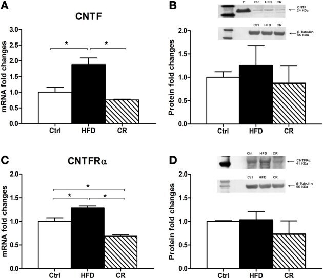
- Figure 2 RT-PCR (A and C) and Western blot (B and D) analyses of CNTF (A and B) and CNTFRalpha (C and D) expression in the hypothalamus of HFD, control (Ctrl) and CR mice. For Western blots the densitometric analysis was normalized to beta-tubulin expression. In (B) recombinant CNTF (P, 30 ng) loaded together with hypothalamic protein extracts served as a positive control. Mean +- SEM. * P < 0.05.
- Submitted by
- Invitrogen Antibodies (provider)
- Main image
- Experimental details
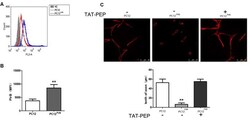
- Figure 2 Overexpression of PirB inhibited neurites outgrowth in PC12 cells, which was reversed by TAT-PEP treatment ( n = 4). Flow cytometry diagram (A) and quantification (B) of PirB expression in PC12 PirB cells (NC means normal PC12 cells without any treatment). (C) Representative images of PC12 cells, PC12 PirB cells with and without treatment of TAT-PEP. Cells were stained with anti-beta-tubulin antibody. The length of axons was measured using fluorescence microscope. ** p < 0.01.

- Submitted by
- Invitrogen Antibodies (provider)
- Main image

- Experimental details
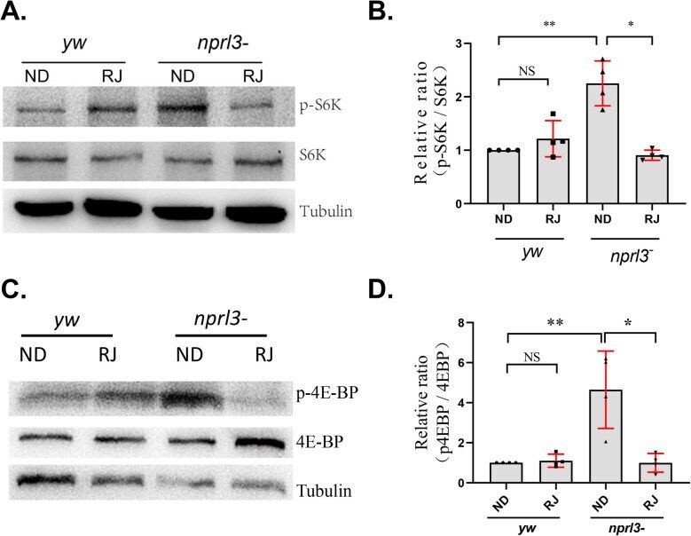
- Fig. 2. RJ feeding decreases TORC1 activity in the nprl3 mutant . yw and nprl3 1 /Df flies were collected from normal food or RJ food. (A) Phosphorylated S6K (top), total S6K (middle), and Tubulin (bottom) in male flies were detected using western blot. (B) The relative intensity of phosphorylated S6K band to total S6K band. (C) Phosphorylated 4E-BP (top), total 4E-BP (middle), and Tubulin (bottom) in male flies were detected using western blot. (D) The relative intensity of phosphorylated 4E-BP band to total 4E-BP band. The ratio of p4E-BP/4E-BP in the yw was set as 1. Data are presented as mean+-s.d. ; values are from four independent experiments; * P
- Submitted by
- Invitrogen Antibodies (provider)
- Main image
- Experimental details
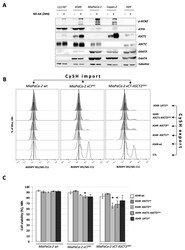
- Figure 4 Genetic disruption of the ASCT2 cysteine transporter affects cooperation between xCT-expressing and xCT KO cells. ( A ). A variety of xCT wt cells (LS174T, A549, MiaPaCa-2, Capan-2 and HDF) were grown in DMEM media +/- amino acids during 24 h and protein content of different cysteine transporters (ASCT1 (SLC1A4), ASCT2 (SLC1A5), EAAT3 (SLC1A1), EAAT4 (SLC1A6) were analyzed by Western blotting. ( B , C ). Lipid hydroperoxide accumulation and cell viability of MiaPaCa-2 xCT KO and xCT-ASCT2 DKO cells (guest cells--CySH import) in control conditions or co-cultured with 6% A549 wt, ASCT1 KO , ASCT2 KO , ASCT1-ASCT2 DKO , or LAT1 KO (host cells--CySH export). All experiments have been performed in triplicate and the representative blots and histograms are shown. Bar graph shows mean +- SEM; n = 3; *, p < 0.05, comparison with WT control group.

- Submitted by
- Invitrogen Antibodies (provider)
- Main image

- Experimental details
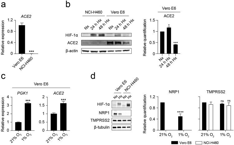
- Figure 2. Hypoxia decreases ACE2 and NRP1 protein levels on Vero E6 cells. ( a ) Relative ACE2 gene expression on Vero E6 and NCI-H460 measured by Q-PCR ( n = 3, unpaired t test). ( b ) (Left) Western blot of HIF-1alpha, ACE2 and beta-actin on NCI-H460 and Vero E6 cells cultured under normoxia (21% O 2 ) or hypoxia (1% O 2 ) for the indicated time points. (Right) Relative quantification of ACE2 protein expression by densitometry ( n = 2, one-way ANOVA). ( c ) Relative gene expression of PGK1 (left) and ACE2 (right) on Vero E6 cells cultured under normoxia or hypoxia for 24 h ( n = 3, unpaired t test). ( d ) (Left) Western blot of HIF-1alpha, NRP1, TMPRSS2 and beta-tubulin on Vero E6 and NCI-H460 cells cultured under normoxia or hypoxia for 48 h. (Right) Relative quantification of NRP1 and TMPRSS2 proteins by densitometry ( n = 3, 2-way ANOVA). Error bars represent SEM and asterisks represent p values (*
- Submitted by
- Invitrogen Antibodies (provider)
- Main image
- Experimental details
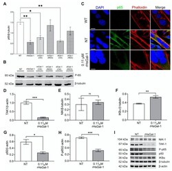
- Figure 2 In vitro treatment with rHsGal-1 modulates inflammatory response through the NF-kappaB pathway. ( A ) Quantification of expression levels of p65 (normalized to beta-tubulin) in 48 h A/J -/- NT or 0.11 muM rHsGal-1 treated myotubes. ( B ) Western blot images showing the p65 expression in NT or 0.11 muM rHsGal-1 48 h A/J -/- treated myotubes. ( C ) Representative images of WT and NT or 0.11 muM rHsGal-1 48 h A/J -/- treated myotubes cultured and immunostained with p65 (green), Phalloidin (red), and DAPI (blue). ( D - H ) Western blot quantification of 48 h NT or 0.11 muM rHsGal-1treated myotubes expressing levels of TAK1 ( D ), NIK ( E ), IKBalpha ( F ), p50 ( G ), and P-p65 ( H ). ( I ) Western blot images of 48 h NT or 0.11 muM rHsGal-1treated myotubes expressing NF-kappaB inflammatory subunits quantified in D-H. n = 3 in each group. A. * p < 0.05 and ** p < 0.01 NT vs. all forms of Gal-1. D-H. * p < 0.05, ** p < 0.01, *** p < 0.001 NT vs. rHsGal-1. Data are represented as SEM.

- Submitted by
- Invitrogen Antibodies (provider)
- Main image
- Experimental details
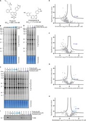
- Target identification using chemical probe and chemoproteomics (A) Chemical structures of probe 4 and OXS008255 5 . EC 50 values for CD11b upregulation are represented as means +- SEM. In-gel fluorescence showing: (B) dose-dependent labelling by probe 4 ; (C) competition of OXS008255 5 and paclitaxel with probe 4 ; (D) competition of inactive analogue OXS007564 6 with probe 4 . Coomassie stain shows equal protein loading on each gel. Volcano plots showing significantly enriched proteins in the pull-down experiment by probe 4 compared to competition with: (E) 1 muM of OXS008255 5 ; (F) 5 muM of OXS008255 5 ; (G) 25 muM of OXS008255 5 ; (H) 25 muM of paclitaxel. For (E)-(H) a student's t-test (FDR = 0.05; S0 = 0.1) was performed between the active probe sample and each DMSO control, and between active probe sample and probe/parent competition samples. Full list of proteins for each volcano plot is available in the supplementary information; (I) Confirmation of tubulin beta chain enrichment by pull-down and immunoblotting. Western blot is cropped, full western blot available in Figure S5 . See also Figure S5 and Tables S3 , S4-S7 , and S8 .

- Submitted by
- Invitrogen Antibodies (provider)
- Main image
- Experimental details
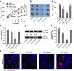
- Figure 6 The antitumour and gene knockdown effects of GNC-siRNA complex in subcutaneous pancreatic tumours. Panc-1 cells were injected into the flank of Balb/c nude mice to form subcutaneous tumours. When the tumours reached about 5 mm in diameter, the animals received peritumoral injections of various formulations every 2 days for six injections. ( a ) Tumour growth curve during the treatments. The black arrows indicated the days of injection. ( b ) Ex vivo tumour image and ( c ) tumour weight at the end of experiment. Scale bar, 1 cm. ( d ) Expression level of NGF mRNA and ( e ) NGF protein level in subcutaneous tumours. ( f ) IF staining of neurites in tumour tissues with various siRNA treatments. Neurites were stained with neurofilament antibody (red), the cell nuclei were stained with 4,6-diamidino-2-phenylindole (DAPI; blue). Scale bars, 20 mum. ( g ) Quantification of neurite density in the subcutaneous tumours by counting the neurite area positive to neurofilament antibody per unit area. Mean+-s.d. ( n =6 per group). Significant difference was from the saline control, * P

- Submitted by
- Invitrogen Antibodies (provider)
- Main image
- Experimental details
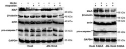
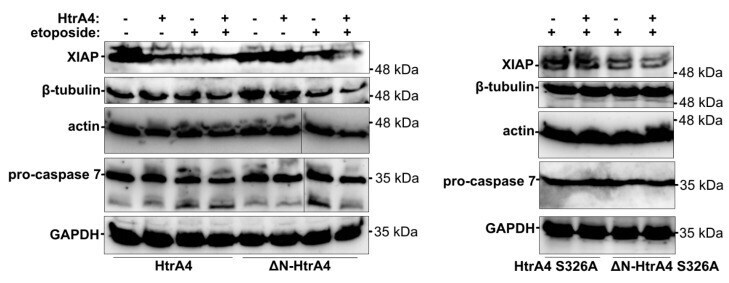
- Figure 9 HtrA4 promotes degradation of XIAP and, less efficiently, of beta-tubulin, actin, and pro-caspase 7. The A549 cells with exogenous HtrA4, DeltaN-HtrA4, or their inactive variants (+) induced by adding doxycycline to the medium were treated with 15 uM etoposide for 48 h. Control cells (-) were incubated with etoposide but without doxycycline. The cells and medium were collected and probed with specific antibodies. Representative blots are presented. Densitometric analyses of the immunoblotting results are shown in Figure S5 .
- Submitted by
- Invitrogen Antibodies (provider)
- Main image

- Experimental details
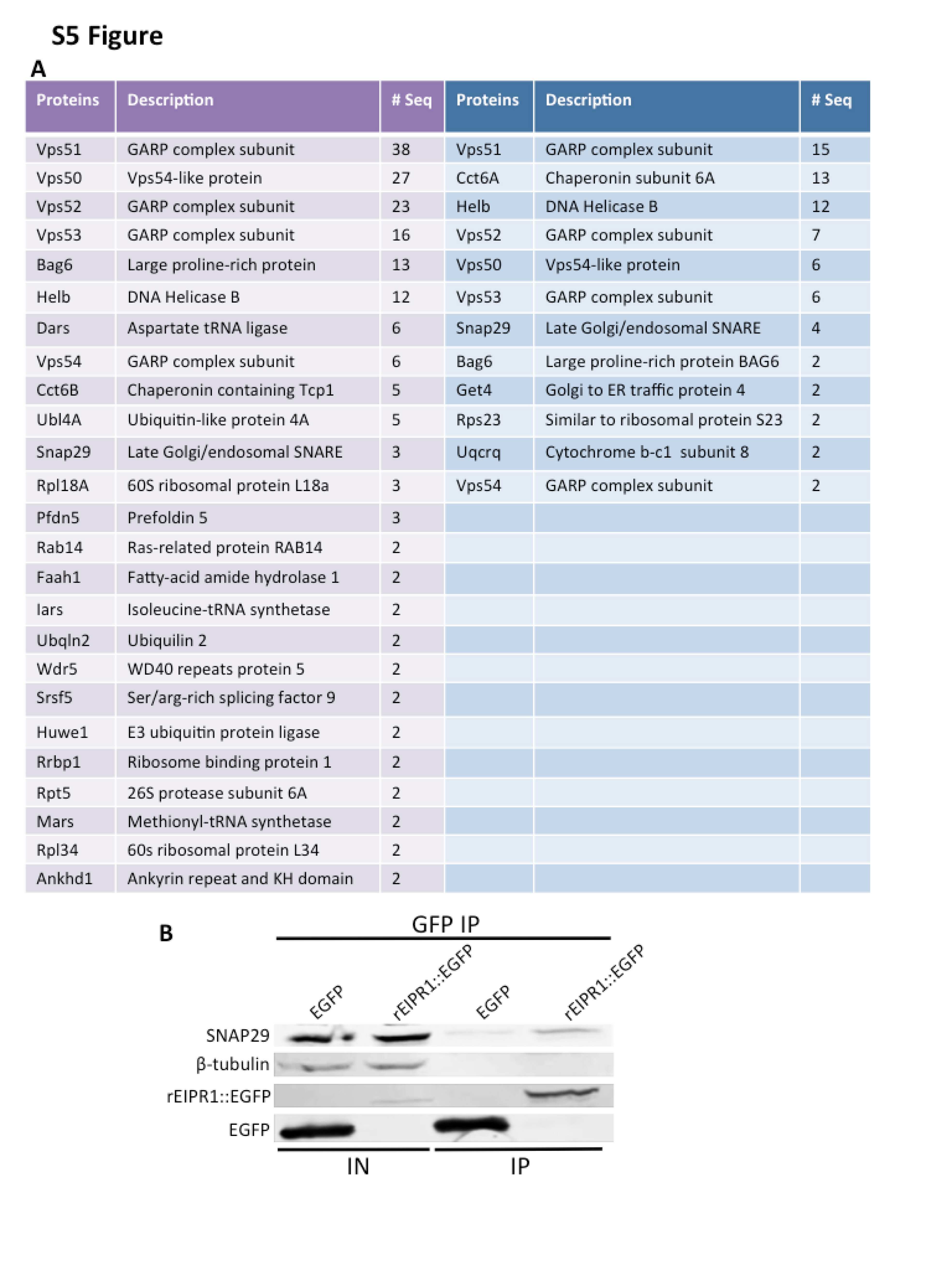
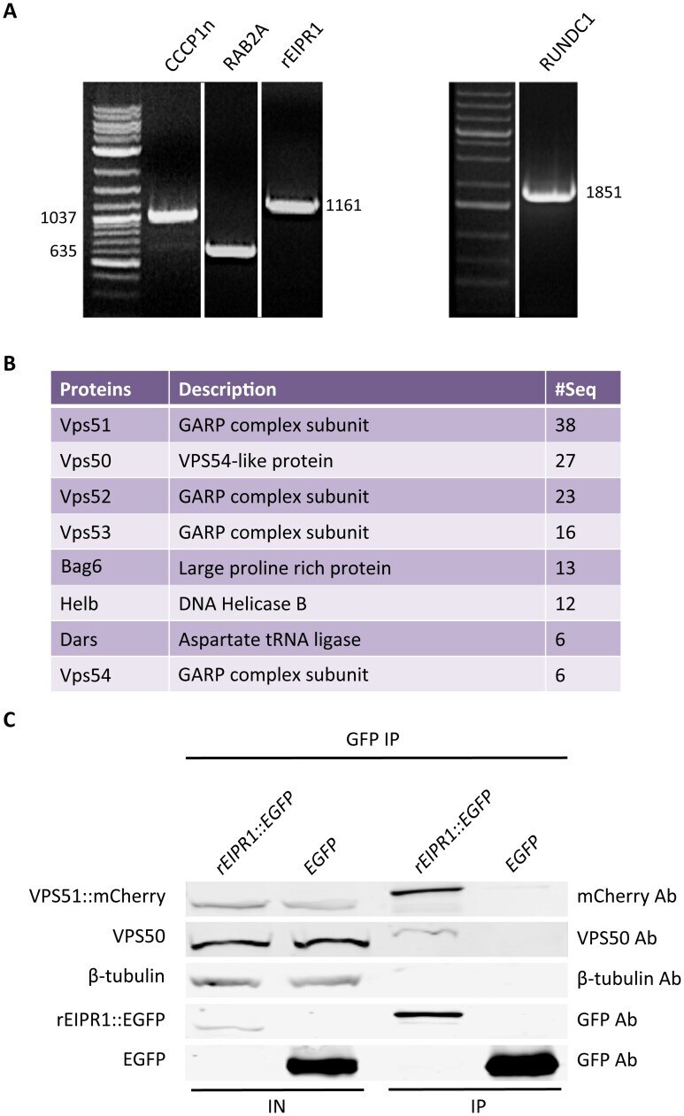
- Fig 4 EIPR-1 interacts with members of the GARP and EARP complexes. (A) The rat ortholog of EIPR-1 is expressed in 832/13 cells. Left and middle: RT-PCR shows that rat orthologs of CCCP-1, RAB-2, EIPR-1 and RUND-1 are expressed in 832/13 cells. All bands are of the predicted size for the full-length cDNA except for CCCP1, which is truncated and corresponds to the N-terminal half of the gene. (B) EIPR1 interacts with members of the GARP and EARP complexes. List of top hits from mass spectrometry of a pulldown of rEIPR1::GFP in 832/13 cells after subtracting hits found in GFP control pulldowns. # seq = number of unique peptides from each protein. All proteins with more than 5 unique peptides are shown from one of two independent experiments. More complete data tables for both experiments are shown in S5A Fig . (C) EIPR1 interacts with VPS51 and VPS50. EGFP-tagged rat EIPR1 or EGFP was coexpressed with mCherry-tagged rat VPS51 in 832/13 cells. Immunoprecipitation of EIPR1::EGFP pulled down VPS51::mCherry and endogenous VPS50. Immunoprecipitation of untagged EGFP did not pull down VPS51::mCherry or VPS50. IN: input; IP: immunoprecipitation.
- Submitted by
- Invitrogen Antibodies (provider)
- Main image
- Experimental details
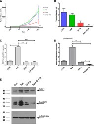
- Figure 5 SI113 sensitizes A2780TC xenografts in immunocompromised mice to paclitaxel. (A) Tumor growth curves of animals treated with vehicle alone (control) SI113, paclitaxel, or both agents together. The data are expressed as mm 3 +- standard error (SE) and were evaluated by one-way ANOVA. (B) The mice were sacrificed 21 days after the beginning of the treatment, and their tumors were excised and weighed. The histogram shows the tumor weight (in grams) of each experimental arm expressed in means +- SEs. The differences among the groups were analyzed by one-way ANOVA. (C, D) Quantitative RT-PCR analysis of SGK1 (left) and RANBP1 (right) mRNA levels in tumors from the above-mentioned four experimental arms. The results (normalized to the HPRT1 levels) are expressed as the average relative fold expression values +- SDs from triplicate experiments and were evaluated by one-way ANOVA. (E) Western blot of proteins extracted from A2780TC-derived xenografts treated as indicated. Cell extracts were separated by SDS-PAGE and detected with SGK1 and RANBP1 antibodies. * P

- Submitted by
- Invitrogen Antibodies (provider)
- Main image
- Experimental details
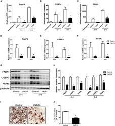
- FIGURE 2 Effect of recombinant irisin and FNDC5 overexpression on adipogenesis. (A-C) 3T3-L1 cells were treated with or without 100 ng/mL irisin from D-2 to D+6 of adipogenesis. Gene expressions of transcription factors were measured by real-time PCR and normalized to 18S rRNA. (D-J) 3T3-L1 preadipocytes were transfected with control or plasmid expressing FNDC5. Two days confluent cells were induced to differentiate and were harvested at day + 5 and day + 8. (D-F) Gene expressions of PPARgamma, CEBPalpha, and FABP4 were analyzed by real-time PCR and normalized to 18S. (G,H) Western blot analysis of transcription factors. (I,J) Representative pictures and quantification of Oil red O staining in FNDC5-overexpressed cells at day 8. * P < 0.05 compared with control at each time-point.

- Submitted by
- Invitrogen Antibodies (provider)
- Main image

- Experimental details
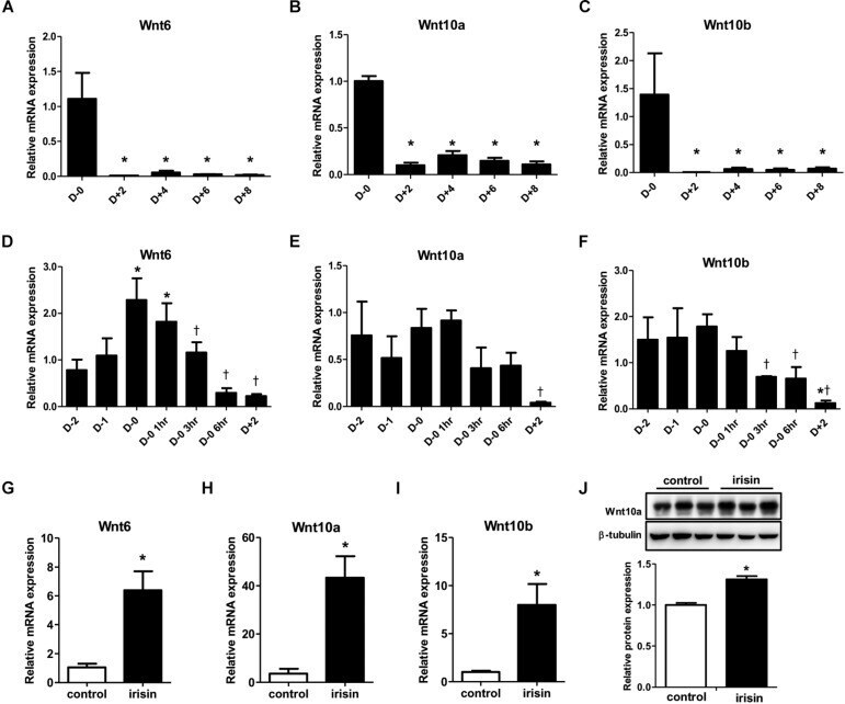
- FIGURE 3 Effect of recombinant irisin treatment on Wnt expression. (A-C) Gene expression analysis of Wnt ligands during 3T3-L1 adipocyte differentiation by real-time PCR. Quantification were normalized to 18S rRNA * P < 0.05 compared with D-0. (D-F) Gene expression of analysis of Wnt ligands at early stage of adipogenesis by real-time PCR. * P < 0.05 compared with D-2, + P < 0.05 compared with D-0. (G-J) 3T3-L1 cells were treated with 100 ng/mL recombinant irisin at D-2. (G-I) Wnt gene expressions were measured by real-time PCR and normalized to 18S rRNA. (J) Protein expression of Wnt10a with or without recombinant irisin treatment were analyzed by Western blot, * P < 0.05 compared with control.
- Submitted by
- Invitrogen Antibodies (provider)
- Main image

- Experimental details
- Fig. 6 PGE2 stimulates PKA/CREB signaling through EP4 to induce sodium influx. a Representative images of sodium indicator (green) analysis pre- and post-PGE2 (20 muM) stimulation for 5 min in primary DRG neurons from EP4 f/f or EP4 -/- mice, indicating sodium influx. Scale bar, 100 mum. Magnification, scale bar, 20 mum. b , c Quantitative analysis of the fluorescent density distribution of the 1st ( b ) and 2nd ( c ) column in ( a ). * p < 0.05, ** p < 0.01 compared with the corresponding pre-treatment group. n = 3 per group. d Western blots of the phosphorylation of PKA and CREB in primary DRG neurons treated with PGE2 (20 muM) for 30 min and PKA inhibitor (H-89, 10 muM) for 60 min. e Quantitative analysis of ( d ). ** p < 0.01 compared with the negative control group from EP4 f/f mice. # p < 0.05, ## p < 0.01 compared with only PGE2 treatment group from EP4 f/f mice n = 3 per group. f First to third row, representative images of immunofluorescent analysis of PKA (red), p-PKA (green) staining, and DAPI (blue) staining of nuclei; 4th to 6th row, representative images of immunofluorescent analysis of CREB (red), p-CREB (green) staining, and DAPI (blue) staining of nuclei pre- and post-PGE2 (20 muM) stimulation combined with H-89 (10 muM) in primary DRG neurons from EP4 f/f or EP4 -/- mice. Scale bar, 100 mum. g Representative images of sodium indicator (green) analysis pre- and post-PGE2 (20 muM) stimulation combined with cAMP, PKA inhibitor (H-89), or siRNA for Na v 1.8 (si-