 Explore
 Validate
 Learn
Learn Western blot
 Immunoprecipitation
Antibody data
- Antibody Data
- Antigen structure
- References [0]
- Comments [0]
- Validations
- Western blot [1]
- Other assay [1]
Submit
Validation data
Reference
Comment
Report error
- Product number
- MA5-15183 - Provider product page

- Provider
- Invitrogen Antibodies
- Product name
- JNK2 Monoclonal Antibody (H.715.7)
- Antibody type
- Monoclonal
- Antigen
- Other
- Description
- It is not recommended to aliquot this antibody.
- Reactivity
- Human, Mouse, Rat, Hamster
- Host
- Rabbit
- Isotype
- IgG
- Antibody clone number
- H.715.7
- Vial size
- 200 µL
- Storage
- -20°C
No comments: Submit comment
Supportive validation
- Submitted by
- Invitrogen Antibodies (provider)
- Main image

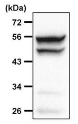
- Experimental details
- Western blot analysis SAPK/JNK was performed by loading 30 µg of THP-1 whole cell lysate per well onto an SDS-PAGE gel. Proteins were transferred to a PVDF membrane and blocked with 5% non-fat dry milk in TBST for 1 hour at room temperature. The membrane was probed with a SAPK/JNK Antibody (Product # MA5-15183) at a dilution of 1:1000 overnight at 4°C, washed in TBST, and probed with an HRP-conjugated goat anti-rabbit IgG secondary antibody at a dilution of 1:40,000 for 1 hour at room temperature. Detection was performed using ECL substrate. Data courtesy of the Innovators Program.

Supportive validation
- Submitted by
- Invitrogen Antibodies (provider)
- Main image
- Experimental details
- Immunoprecipitation of SAPK/JNK was performed on HEK293T cells. Antigen-antibody complexes were formed by incubating 500 µg of HEK293T whole cell lysate (in 500 µL volume) with 5 µL of a SAPK/JNK monoclonal antibody (Product # MA5-15183) overnight at 4°C. The immune complexes were captured on 30 µL of protein G agarose, washed extensively, and eluted with 6X Laemmli buffer. Samples were resolved on a 10% SDS-PAGE gel, transferred to a PVDF membrane, and blocked with 5% milk in TBST for 1 hour at room temperature. The membrane was probed with a SAPK/JNK monoclonal antibody (Product # MA5-15183) at a dilution of 1:1000 overnight at 4°C, washed in TBST, and probed with an HRP-conjugated mouse anti-rabbit light chain-specific secondary antibody at a dilution of 1:40,000 for 1 hour at room temperature. Chemiluminescent detection was performed using ECL substrate. Data courtesy of the Innovators Program.
