Explore
 Validate
 Learn
Learn Western blot
 Immunohistochemistry
Antibody data
- Antibody Data
- Antigen structure
- References [1]
- Comments [0]
- Validations
- Western blot [2]
- Immunocytochemistry [1]
Submit
Validation data
Reference
Comment
Report error
- Product number
- PA1-9046 - Provider product page

- Provider
- Invitrogen Antibodies
- Product name
- GAPDH Polyclonal Antibody
- Antibody type
- Polyclonal
- Antigen
- Synthetic peptide
- Description
- This antibody is predicted to react with canine based on sequence homology.
- Reactivity
- Human, Rat
- Host
- Goat
- Isotype
- IgG
- Vial size
- 100 µg
- Concentration
- 0.5 mg/mL
- Storage
- -20° C, Avoid Freeze/Thaw Cycles
Submitted references Nuclear Envelope Transmembrane Proteins in Myotonic Dystrophy Type 1.
Hintze S, Knaier L, Limmer S, Schoser B, Meinke P
Frontiers in physiology 2018;9:1532
Frontiers in physiology 2018;9:1532
No comments: Submit comment
Supportive validation
- Submitted by
- Invitrogen Antibodies (provider)
- Main image
- Experimental details
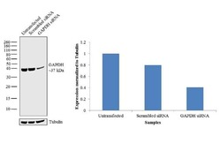
- Knockdown of GAPDH was achieved by transfecting HeLa cells with GAPDH specific validated siRNAs (Silencer® select Product # s5572, s5573). Western blot analysis (Fig. a) was performed using whole cell extracts from the GAPDH knockdown cells (lane 3), non-specific scrambled siRNA transfected cells (lane 2) and untransfected cells (lane 1). The blot was probed with GAPDH Polyclonal Antibody (Product # PA1-9046, 1 µg/mL) and Rabbit anti-Goat IgG (H+L) Superclonal™ Secondary Antibody, HRP conjugate (Product # A27014, 0.25 µg/mL, 1:4000 dilution). Densitometric analysis of this western blot is shown in histogram (Fig. b). Decrease in signal upon siRNA mediated knock down confirms that antibody is specific to GAPDH.

- Submitted by
- Invitrogen Antibodies (provider)
- Main image
- Experimental details
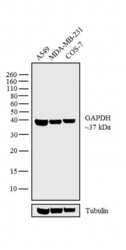
- Western blot analysis was performed on whole cell extracts (30 µg lysate) of A549 (Lane 1), MDA-MB-231 (Lane 2) and COS-7 (Lane 3). The blot was probed with Anti-GAPDH Polyclonal Antibody (Product # PA1-9046, 1 µg/mL) and detected by chemiluminescence using Rabbit anti-Goat IgG (H+L) Superclonal™ Secondary Antibody, HRP conjugate (Product # A27014, 0.25 µg/mL, 1:4000 dilution). A 37 kDa band corresponding to GAPDH was observed across the cell lines tested.

Supportive validation
- Submitted by
- Invitrogen Antibodies (provider)
- Main image

- Experimental details
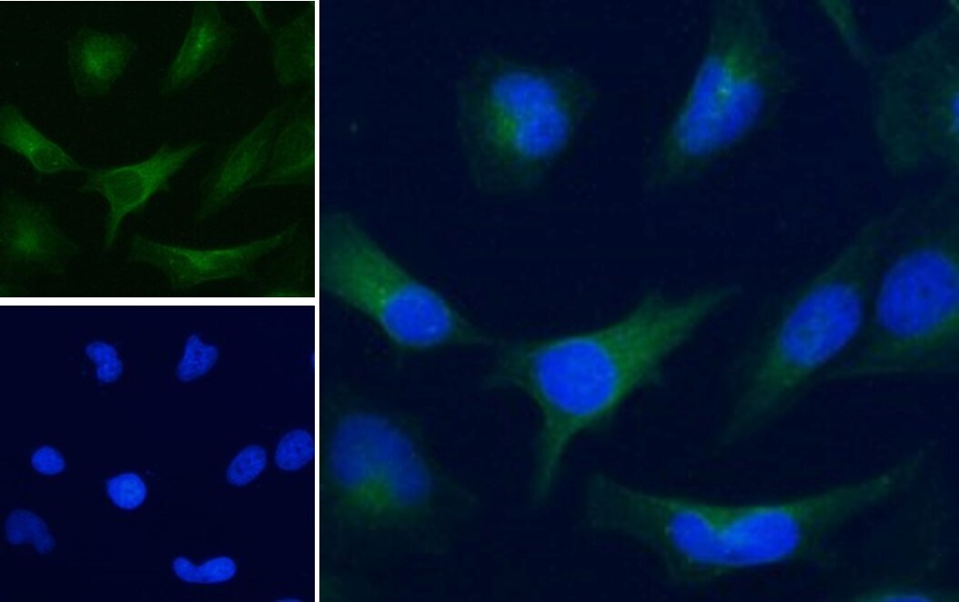
- Immunofluorescence analysis of GAPDH in HeLa cells using a GAPDH monoclonal antibody (Product # PA1-9046) at 5 µg/mL for1hr. The cells were paraformaldehyde fixed and permeabilized with 0.15% Triton. Primary incubation was followed by Alexa Fluor 488 secondary antibody (1 µg/mL) showing cytoplasmic staining. The nuclear stain is DAPI (blue).