 Explore
 Validate
 Learn
Learn Western blot
Antibody data
- Antibody Data
- Antigen structure
- References [0]
- Comments [0]
- Validations
- Western blot [1]
- Other assay [6]
Submit
Validation data
Reference
Comment
Report error
- Product number
- PA1-987-HRP - Provider product page

- Provider
- Invitrogen Antibodies
- Product name
- GAPDH Polyclonal Antibody, HRP
- Antibody type
- Polyclonal
- Antigen
- Synthetic peptide
- Description
- By Western blot, PA1-987-HRP detects GAPDH at ~37kD band in human, mouse, and rat whole cell lysates.
- Reactivity
- Human, Mouse, Rat
- Host
- Rabbit
- Conjugate
- Horseradish Peroxidase
- Isotype
- IgG
- Vial size
- 50 µL
- Concentration
- 1 mg/mL
- Storage
- 4° C, do not freeze
No comments: Submit comment
Supportive validation
- Submitted by
- Invitrogen Antibodies (provider)
- Main image

- Experimental details
- Western blot analysis of GAPDH was performed by loading various whole cell lysates onto a 4-20% Tris-HCl polyacrylamide gel. Proteins were transferred to a PVDF membrane and blocked with 5% BSA/TBST for at least 1 hour. Membranes were probed with GAPDH polyclonal antibody (Product # PA1-987-HRP) at a dilution of 1:500 for 1 hour at room temperature on a rocking platform and washed in TBS-0.1% Tween-20. Chemiluminescent detection was performed using Super Signal West Pico (Product # 34080).
- Conjugate
- Horseradish Peroxidase

Supportive validation
- Submitted by
- Invitrogen Antibodies (provider)
- Main image

- Experimental details
- NULL
- Conjugate
- Horseradish Peroxidase

- Submitted by
- Invitrogen Antibodies (provider)
- Main image

- Experimental details
- Figure 1 Detection of AOC3 overexpression in RIMECs by adenovirus. (a) AOC3 mRNA expression in RIMECs as determined by qRT-PCR. GAPDH was used as an internal control. (b) SSAO protein expression in RIMECs was determined by Western-Blot. GAPDH was used as an internal control (* P < 0.05, ** P < 0.01, and *** P < 0.001).
- Conjugate
- Horseradish Peroxidase

- Submitted by
- Invitrogen Antibodies (provider)
- Main image

- Experimental details
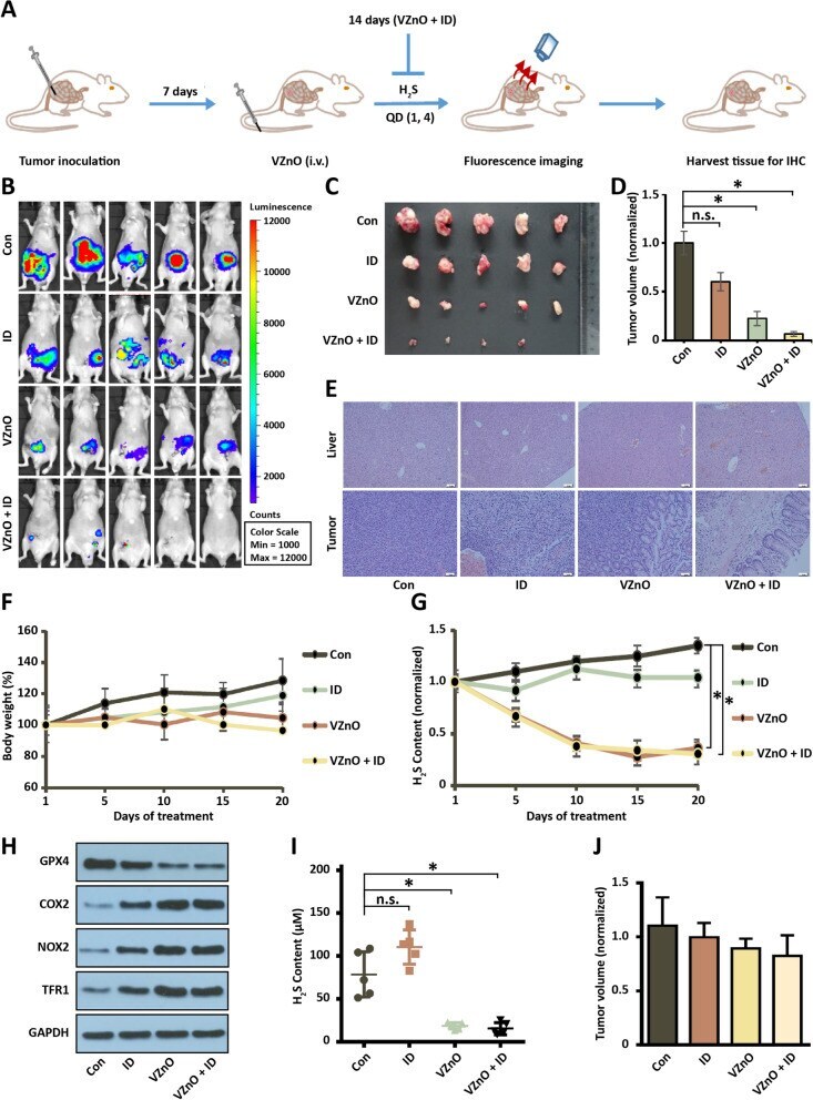
- Fig. 5 Anti-tumor activity of VZnO in orthotopic CRC model. A Experimental timeline of the orthotopic CRC model. B Bioluminescence imaging of orthotopic colon cancer mouse injected with different treatments on day 14. C , D Photograph and volume of the tumors with different treatments on day 14. E Representative H&E stained of liver and tumor tissue on day 14. F Body weights of orthotopic colon cancer model. G The continuously H 2 S production in mouse model. H Representative GPX4, COX2, NOX1 and TFR1 protein expression by Western Blot in tumor on day 14. Membranes were re-probed for GAPDH expression to show that similar amounts of protein were loaded in each lane. I) H 2 S content measured on day 14. J The volume of the tumors in the orthotopic breast model on day 14. (* P < 0.05), (n.s. : No significant difference, P value > 0.05)
- Conjugate
- Horseradish Peroxidase
- Submitted by
- Invitrogen Antibodies (provider)
- Main image
- Experimental details
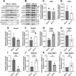
- Protein expression in murine and human primary fibroblasts. ( A ) Western blots for JAG1, DUSP7, ITGB3 and MAP2K1, each with GAPDH, of three independent replicates (each band represents a pool of three technical triplicates) in PMFs. ( B ) Western blots for JAG1, DUSP7, ITGB3 and MAP2K1, with GAPDH of three independent replicates (each band represents a pool of three technical triplicates) in HUAFs. ( C - L ) Quantifications of protein band intensities, normalized to GAPDH. Data are represented as mean +- SEM. * * P < 0.01, compared with GM-ctrl.
- Conjugate
- Horseradish Peroxidase

- Submitted by
- Invitrogen Antibodies (provider)
- Main image

- Experimental details
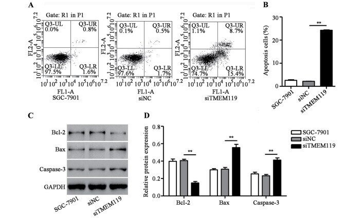
- Figure 3. TMEM119 silencing induced SGC-7901 cell apoptosis. After transfection of SGC-7901 cells with TMEM119-siRNA (siTMEM119), cell apoptosis was measured by (A and B) flow cytometry assay and the expression of Bcl-2, Bax as well as caspase-3 was measured by (C and D) western blot analysis. **P
- Conjugate
- Horseradish Peroxidase
- Submitted by
- Invitrogen Antibodies (provider)
- Main image

- Experimental details
- Figure 3. Experimental validation of miR-183-5p.1 as a TPM1 regulator. (A) miR-183-5p.1 binding with the 3'UTR of TPM1 predicted by TargetScan and coding sequence of miR-183-5p.1 and TPM1. The luciferase activities of WT-TPM1 mRNAs in GC (B) AGS and (J) HGC-27 cells were significantly enhanced by transfection with miR-183-5p.1 inhibitors and markedly decreased in (F) AGS and (O) HGC-27 cells by miR-183-5p.1 mimics following the Luciferase reporter assay. TPM1 expression was markedly increased in (C-E) AGS and (K-N) HGC-27 cells by miR-183-5p.1 inhibitors and significantly decreased in (G-I) AGS and (P-R) HGC-27 cells by miR-183-5p.1 mimics as revealed by reverse transcription-quantitative PCR and western blotting. GAPDH served as the internal control. Results are presented as the mean +- standard error of the mean (n=3). *P
- Conjugate
- Horseradish Peroxidase
