 Explore
 Validate
 Learn
Learn Western blot
Antibody data
- Antibody Data
- Antigen structure
- References [0]
- Comments [0]
- Validations
- Western blot [2]
- Other assay [4]
Submit
Validation data
Reference
Comment
Report error
- Product number
- 14-9523-82 - Provider product page

- Provider
- Invitrogen Antibodies
- Product name
- GAPDH Monoclonal Antibody (FF26A), eBioscience™
- Antibody type
- Monoclonal
- Antigen
- Other
- Description
- Description: This FF26A monoclonal antibody reacts with human glyceraldehyde-3-phosphate dehydrogenase (GAPDH), a key component of glycolysis. This 36 kDa enzyme is constitutively expressed in all tissues albeit at different levels. Measurement of GAPDH levels is often performed as a control for protein loading in experiments assessing differential protein expression. The FF26A antibody is crossreactive to mouse GAPDH. Applications Reported: This FF26A antibody has been reported for use in western blotting. Applications Tested: This FF26A antibody has been tested by immunoblotting of cell lysates. This can be used at less than or equal to 1 µg/mL. It is recommended that the antibody be carefully titrated for optimal performance in the assay of interest. Purity: Greater than 90%, as determined by SDS-PAGE. Aggregation: Less than 10%, as determined by HPLC. Filtration: 0.2 µm post-manufacturing filtered.
- Reactivity
- Human, Mouse
- Host
- Mouse
- Isotype
- IgG
- Antibody clone number
- FF26A
- Vial size
- 100 µg
- Concentration
- 0.5 mg/mL
- Storage
- 4° C
No comments: Submit comment
Supportive validation
- Submitted by
- Invitrogen Antibodies (provider)
- Main image

- Experimental details
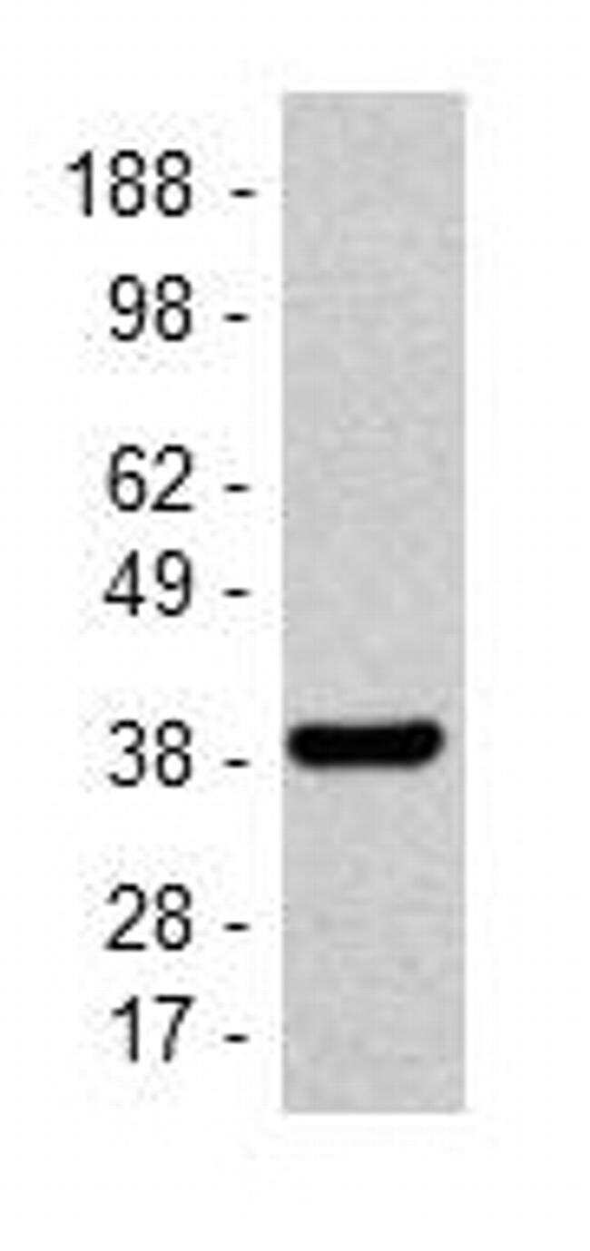
- Immunoblotting of reduced HeLa cells using 5 µg/mL of Anti-Human GAPDH Purified antibody.Bands were visualized using Anti-Mouse IgG HRP.
- Submitted by
- Invitrogen Antibodies (provider)
- Main image
- Experimental details
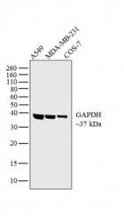
- Western blot analysis was performed on whole cell extracts (30 µg lysate) of A549 (Lane 1), MDA-MB-231 (Lane 2) and COS-7 (Lane 3). The blot was probed with Anti-GAPDH Monoclonal Antibody (Product # 14-9523-80, 1 µg/mL) and detected by chemiluminescence using Goat anti-Mouse IgG (H+L) Superclonal™ Secondary Antibody, HRP conjugate (Product # A28177, 0.25 µg/mL, 1:4000 dilution). A 37 kDa band corresponding to GAPDH was observed across the cell lines tested.

Supportive validation
- Submitted by
- Invitrogen Antibodies (provider)
- Main image

- Experimental details
- NULL

- Submitted by
- Invitrogen Antibodies (provider)
- Main image

- Experimental details
- Figure 3. Expression of Arg1, iNOS and IL-10 in lung IMs of OVA-induced asthmatic mice. Lung IMs from OVA-induced asthmatic mice and PBS-treated control mice were sorted 24 h after the final OVA challenge (day 27 post-model establishment; 100 ug in 50 ul PBS) and (A) western blot analysis was used to measure Arg1, IL-10 and iNOS protein expression. GAPDH was used as a protein loading control and images are representative of 6 mice in each group. ODs of (B) Arg1, (C) iNOS and (D) IL-10 levels normalized to GAPDH. Data are presented as the mean +- standard deviation, n=6. **P

- Submitted by
- Invitrogen Antibodies (provider)
- Main image

- Experimental details
- Fig 6 Normalizing beta-arrestin-dependent signaling of CXCR4 1013 partially restores the productive HPV life cycle. (A) Western blots (left) and densitometric analyses (right) showing relative levels of HPV18-E2 protein in HPV18-positive CXCR4 wt , CXCR4 1013 , and CXCR4 1013&DeltaSHSK raft cultures. E2 protein levels were normalized to GAPDH protein levels and arbitrarily set at 1 for CXCR4 wt rafts (staining controls are shown in S5 Fig ). Values are means +- SEM. **p < 0.01 and ***p < 0.001. HPV18-positive CXCR4 1013&DeltaSHSK raft sections stained for HPV18-E4 (B), HPV18-L1 (C), and Ki-67 (D) expression by immunohistochemical staining. Images are representative of three independent experiments. Scale bars = 100 mum, inset scale bar = 10 mum.

- Submitted by
- Invitrogen Antibodies (provider)
- Main image

- Experimental details
- Figure 3. Melatonin Treatment Significantly Improves Cell Survival via the Activation of PI3K/Akt Pathway After Ischemic Stroke In Vitro and In Vivo . A-F: In vitro , N2a cells were treated with 25 nM melatonin for 24 hours after OGD in the presence or absence of wortmannin. A: Cell survival. B: Evaluation of the purity of the cell membrane and cytoplasm isolation of N2a cells. Western blots analysis of the different protein markers sodium potassium ATPase (plasma membrane), GRP94 (endoplasmic reticulum), Ku86 (nuclear) and GAPDH (cytoplasm). C: Representative western blots of p-Akt and Akt in the membrane and cytoplasm fractions. Quantification of western blots data of Akt (D), p-Akt expression (E), p-Akt/Akt ratio (F), respectively. The data resulted from Figure 3C . Values are mean +- SEM. *** p < 0.001 vs . vehicle; ## p < 0.01, ### p < 0.001 vs . vehicle plus OGD; ++ p < 0.05, +++ p < 0.01 vs . vehicle plus OGD plus 25 nM melatonin. One-way ANOVA followed by Bonferroni post hoc test. Samples were collected from three independent experiments, each performed in duplicate. G-J: In vivo , mice were subjected to dMCAO and treated with 20 mg/kg melatonin at 72 hours after ischemic stroke. G: Representative western blots of p-Akt and Akt in mice. H: Quantification of Akt, p-Akt expression, and p-Akt/Akt ratio. The data resulted from Figure 3G . I: Representative co-immunostaining of p-Akt (red) and NeuN (green). Nuclei were labeled with DAPI (blue) in each group. Scale bar=50 u
